Harris’s hawk diagnosed with fatal herpesvirus infection at necropsy
Erin Edwards, DVM, MS, DACVP
A 6-month-old Harris’s hawk was necropsied at the Texas A&M Veterinary Medical Diagnostic Laboratory (TVMDL) in College Station and was diagnosed with herpesvirus infection as the cause of death. This hawk was used as an abatement bird and had been previously diagnosed with and treated for oral trichomoniasis. The patient passed away and was submitted for necropsy to search for any underlying diseases that may have predisposed the bird to Trichomonas infection.
At necropsy, oral lesions consistent with trichomoniasis were confirmed. These lesions also extended throughout the esophagus and crop. More notably, the spleen in this hawk was enlarged and was mottled white and tan, indicating a necrotizing splenitis (see Figure 1). Histopathology examination confirmed the presence of large necrotic areas in the spleen. Additionally, there were several scattered cells with intranuclear viral inclusion bodies. Similar inclusion bodies were found in the liver (Figure 2) surrounding large areas of necrosis and were also seen in the oral cavity, esophagus, and crop surrounding ulcerated areas. The lesion distribution and appearance of the viral inclusion bodies led to a diagnosis of herpesvirus infection.
Though few cases of falcon herpesviruses have been identified, the majority of hawks and other birds of prey are infected with columbid herpesvirus 1, a pigeon herpesvirus. Raptors are infected with this virus following ingestion of infected pigeon prey. Due to this hawk’s abatement occupation and known exposure to pigeon prey, the herpesvirus in this case was presumed to be columbid herpesvirus 1.
Trichomoniasis is also a common ailment of raptors acquired through ingestion of pigeon prey. This disease is caused by a protozoal parasite, Trichomonas gallinae. In pigeons the disease is known as canker and in birds of prey it is known as frounce. In this bird, the oral, esophageal, and crop lesions that resembled trichomoniasis grossly were histologically attributed to herpesvirus infection. No obvious T. gallinae organisms were seen histologically. These organisms can be difficult to find histologically, though, and a secondary co-infection could not be excluded. This case emphasizes that herpesvirus should be considered a differential diagnosis for oral cavity lesions that resemble trichomoniasis. Other diseases that can cause similar gross lesions include, but are not limited to, avian poxvirus (wet pox), candidiasis, capillariasis, vitamin A deficiency, and, less commonly, salmonellosis.
For more information about this case, contact Veterinary Pathologist Dr. Erin Edwards. To learn more about TVMDL’s test catalog, visit tvmdl.tamu.edu or call 1.888.646.5623.

Figure 2: Histolopathology of the liver showing large areas of necrosis with frequent intranuclear herpesvirus inclusion bodies within hepatocytes. A few of the inclusion bodies are indicated with arrows.
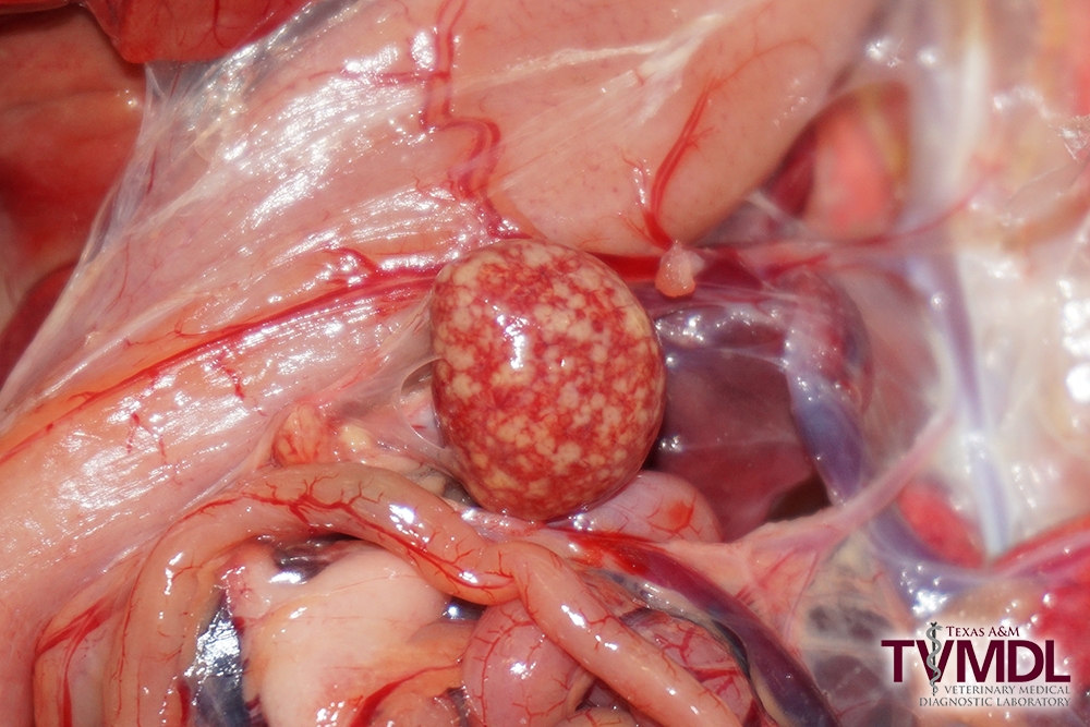
Figure 1: Photograph of the spleen from a Harris’s Hawk with necrotizing splenitis due to herpesvirus infection.