Cytauxzoon felis infection in two cats
Emma Jones and Julie Piccione, DVM, MS, DACVP
An outdoor 5-year-old domestic medium hair cat from Gainesville, Texas was presented to a veterinarian for decreased appetite and thirst of one-day duration. The cat had disappeared a few days prior to examination. Freshly prepared blood smears were submitted for a hemoparasite examination at TVMDL. Examination of the blood smear from the cat revealed schizont-laden macrophages in the feathered edge, as well as round “signet ring” piroplasms in the red blood cells.
A 1-year-old domestic long hair cat was presented to a veterinarian in Crockett, Texas. Whole blood and freshly prepared blood smears were submitted to TVMDL with minimal clinical history available. A complete blood count with hemoparasite exam was performed. Both schizont-laden macrophages and piroplasms were seen in the blood smears of this cat as well. This cat had leukopenia and thrombocytopenia with mild clumping of the platelets.
Cytauxzoon felis typically affects outdoor adult cats in rural or semi-rural areas in most midwestern and southern states. Cases tend to peak in early spring. Cats can present with reduced appetite, lethargy, icterus, pallor, organomegaly, and fever. Pancytopenia can be seen on CBC testing. Chemistry results usually reveal hyperbilirubinemia, hyperglycemia, and hypoproteinemia. The natural reservoir of Cytauxzoon felis is the bobcat. Cytauxzoon is transmitted by the Amblyomma americanum (lone star tick) or Dermacentor variabilis (American dog tick). Once Cytauxzoon merozoites are in the tick, they mature into infective sporozoites in the salivary glands, where they are passed on to domestic cats during feeding. Sporozoites enter mononuclear cells and undergo a form of asexual reproduction (schizogony) where they are released as merozoites that are then taken up by the red blood cells.
As mentioned, schizont-laden macrophages and piroplasms seen on the blood smear of an infected cat can be diagnostic for the disease; however, circulating piroplasms are often rare and may not be seen in early infection. Schizont-laden macrophages are even rarer in circulation/blood smear examination. PCR testing can be performed to catch early infections and to potentially differentiate between other protozoan organisms.
The disease in domestic cats typically progresses rapidly and death within 36 hours is common; however, treatment with supportive care and anti-protozoal drugs have been successful. Additionally, subclinical infections have been recognized in the last few years. Tick control and prophylaxis are the best ways to provide protection for domestic cats.
For more information on TVMDL’s clinical pathology testing, visit tvmdl.tamu.edu or call 1.888.646.5623.
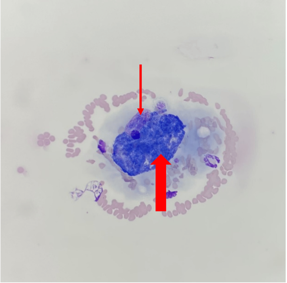
Large schizont-laden mononuclear cell (macrophage) in the feathered edge of a blood smear from a cat. The nucleus of the cell is identified by the thin red arrow. The thick red arrow shows the schizonts. The cell is entrapped in a platelet clump.
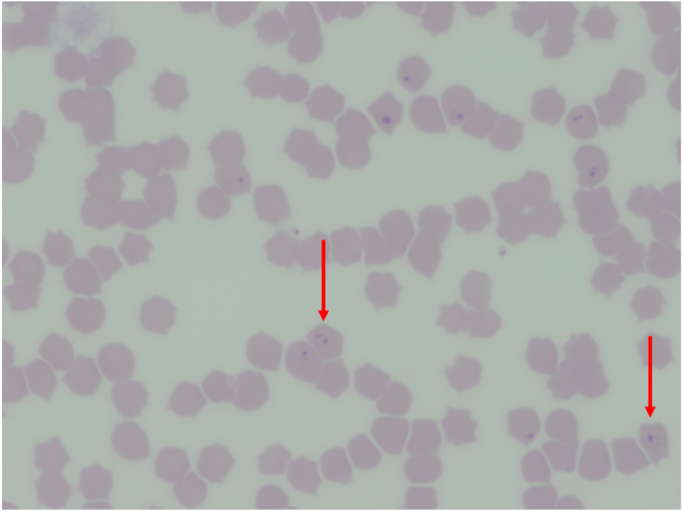
Numerous piroplasms consistent with Cytauxzoon felis within RBCs of a cat.