Aspergillosis diagnosed in 5-month-old chicken
By Martin Ficken, DVM, PhD
A 5-month-old female chicken with a history of dyspnea was presented to the Texas A&M Veterinary Medical Diagnostic Laboratory (TVMDL) in Gonzales for necropsy. Duration of illness was approximately one week. This bird was the only one that had died out of a flock of four.
Upon examination, the bird weighed 692 grams and had moderate breast muscle atrophy. Numerous 1-3 mm, round, cream-colored nodules were present in the anterior and posterior air sacs and were scattered throughout the parenchyma of both lungs (Figure 1).
The right primary bronchus, immediately distal to the syrinx, was completely occluded by caseous inflammatory exudate (Figure 2).
No other significant lesions were observed in any other tissues or organ systems.
Evaluation led to the diagnosis of aspergillosis.
Aspergillosis is a fungal disease caused by infection with the genus Aspergillus, which is composed of approximately 180 species. Manifestations of aspergillosis depend on which organs or systems are involved and whether the infection is localized or disseminated. Aspergillosis in birds usually affects the lower pulmonary system with florid lesions in air sacs and lungs. In young poultry the disease is referred to as brooder pneumonia, “asper,” or fungal pneumonia. Less common manifestations relate to infections of the eye, brain, skin, joints, and viscera.
Aspergillosis is not transmissible from bird to bird but is acquired from environmental exposure. Disturbances of soil or movement of hay, compost, or litter can produce aerosols that provide for respiratory exposure to conidia. Fresh litter, especially if it has been wet previously and contaminated with Aspergillus fumigatus, is associated with outbreaks of aspergillosis. Infection can also occur in hatcheries within incubators, hatchers, incubator rooms, and intake ducts.
Clinical signs can range from subtle to severe. Dyspnea, gasping, and accelerated breathing may be present. Somnolence, inappetence, emaciation, increased thirst, pyrexia, torticollis, and loss of equilibrium have also been described.
Gross lesions consist of small, white, caseous nodules scattered throughout the lung tissue, usually accompanied by similar-sized caseous plaques on thickened air sac membranes. Caseous exudate can also be observed at the level of the syrinx and within the bronchi. The organism can spread systemically to involve the eyes, brain, and other organs.
To learn more about this case, contact Gonzales Resident Director Dr. Martin Ficken. For more information about TVMDL’s test offerings, visit tvmdl.tamu.edu or call 1.888.646.5623.
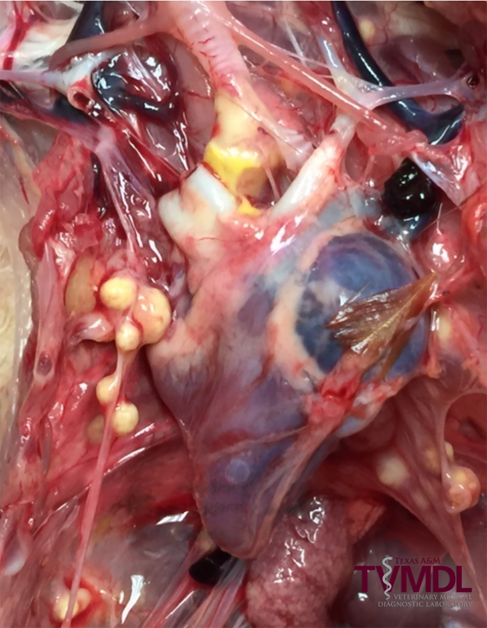
Figure 1. Cream-colored fungal nodules in air sacs.
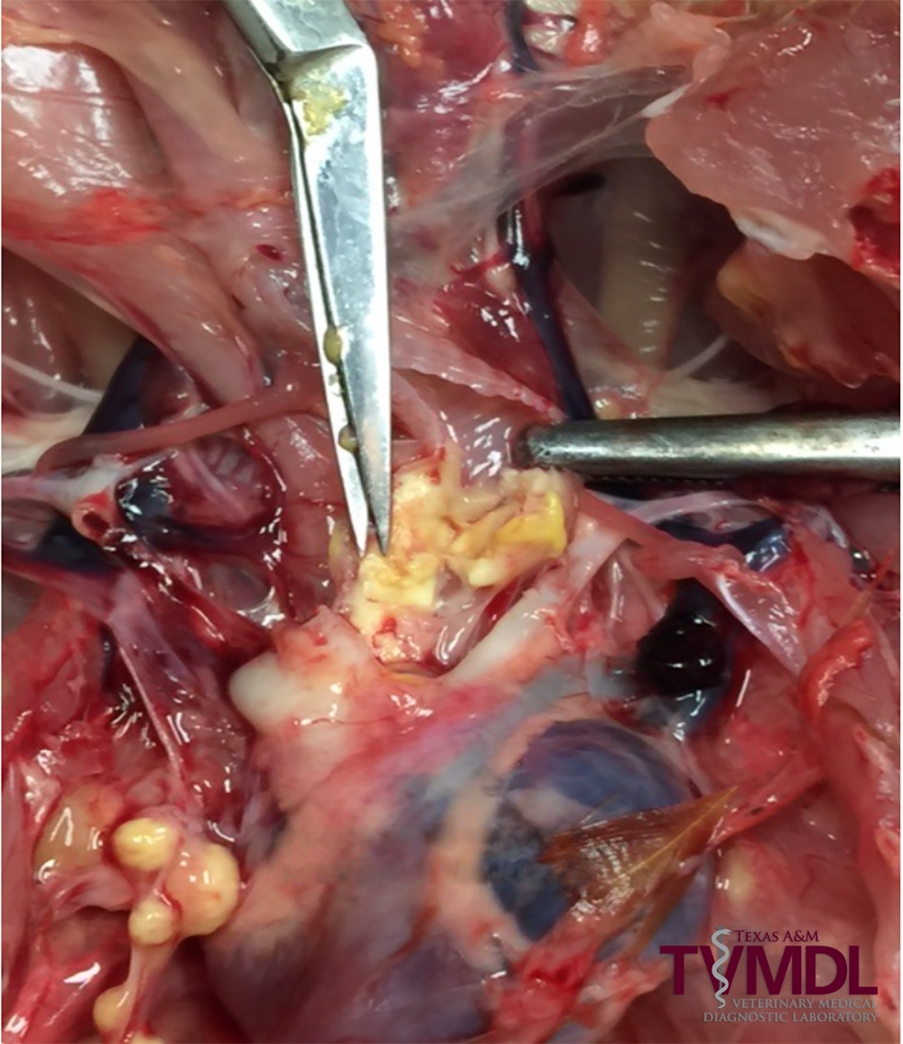
Figure 2. Exudate filled right bronchus.