Disseminated histoplasmosis in a domestic feline
By Amy K. Swinford, DVM, MS, DACVM and Barbara Lewis, DVM, PhD, DACVP
Bone biopsy specimens taken from an adult male castrated domestic feline were submitted to the Texas A&M Veterinary Medical Diagnostic Laboratory (TVMDL) for bacterial and fungal culture and histopathologic examination. The referring veterinarian stated that radiographs of the right hock and right radius and ulna indicated changes consistent with possible osteomyelitis or neoplasia. There were no other radiographic findings, and blood work was within normal limits, but the cat had a loss of appetite and was behaving abnormally.
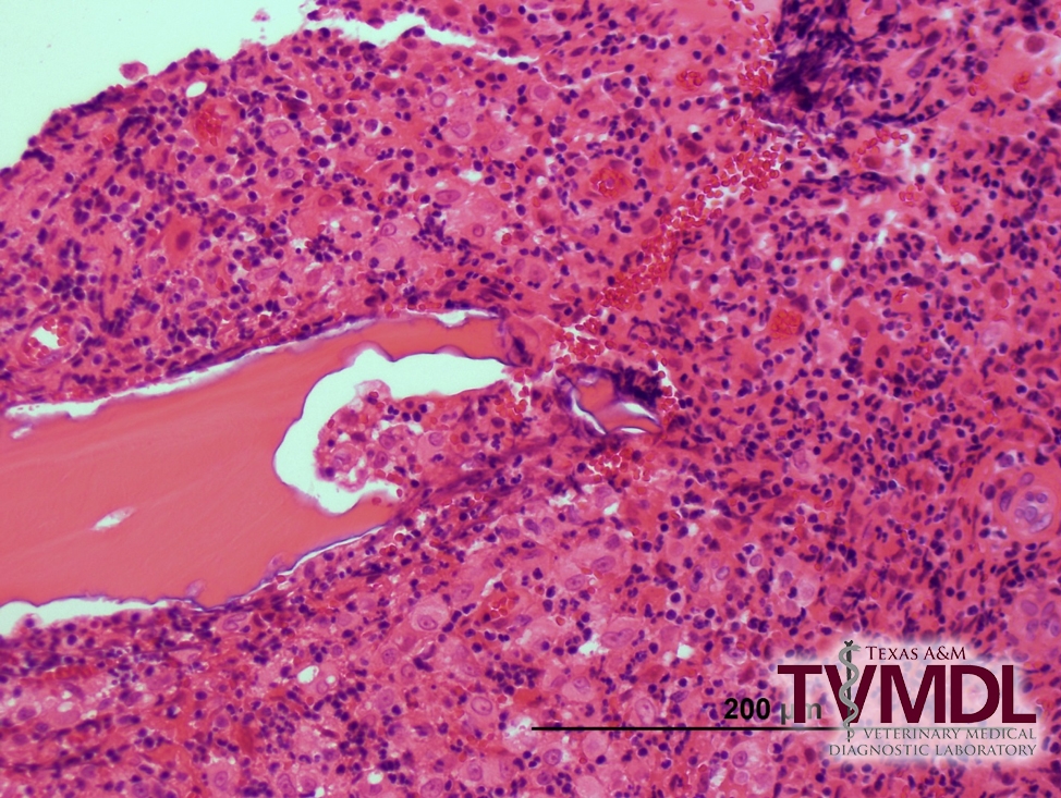
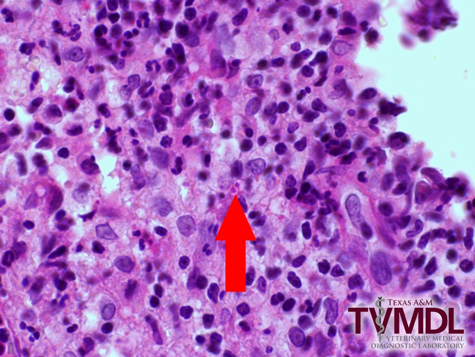
The several sections of bone that were examined by H&E staining revealed pyogranulomatous osteomyelitis, with areas of bone resorption and remodeling (Figure 1). Because the lesion appeared consistent with a systemic fungal infection, additional slides were stained with GMS and PAS (Figure 2) to look for fungal elements. These special stains revealed very low numbers of 1-2 um diameter yeasts with a clear halo, consistent with Histoplasma capsulatum.
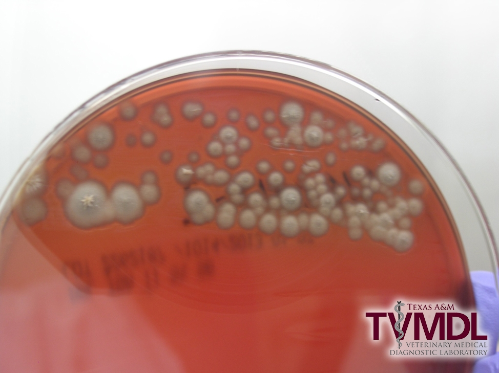
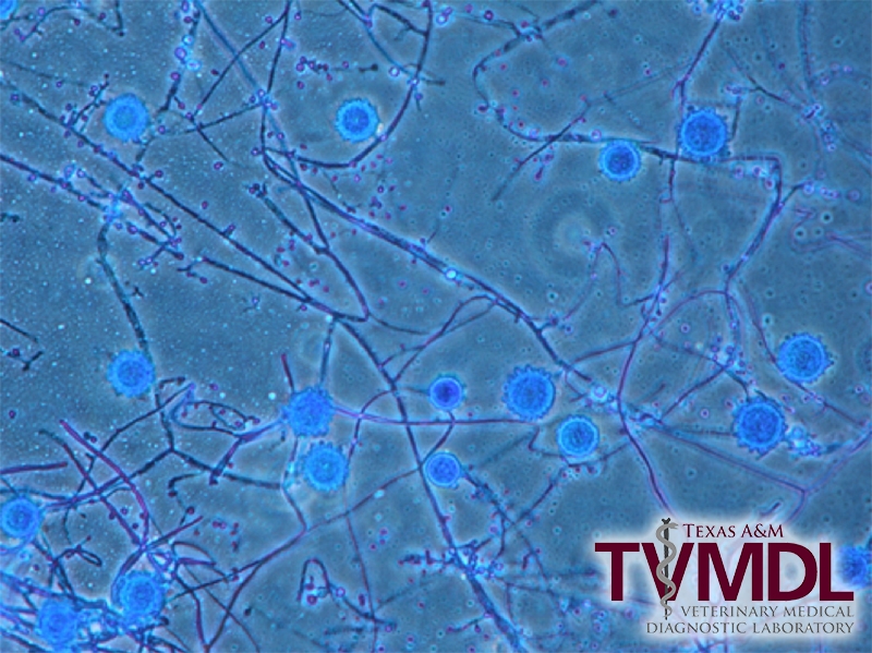
While no bacteria were isolated from bone biopsy samples following extended incubation, tiny fungal colonies appeared after only three days of incubation of the blood agar and Mycobiotic agar plates. Several weeks of incubation yielded large numbers of a pure culture of white, filamentous colonies. The colony characteristics (Figure 3) and microscopic morphology (Figure 4) were consistent with Histoplasma capsulatum, and the identity of the fungal isolate was confirmed with molecular sequence analysis. Upon diagnosis, the cat was placed on itraconazole, but the final clinical outcome was not obtained by TVMDL. The prognosis for disseminated histoplasmosis can be favorable, depending on the severity and sites of infection.
While histoplasmosis has been diagnosed in a wide variety of domestic and exotic animal species, it is most commonly diagnosed at TVMDL in canines and felines. Histoplasmosis is a zoonotic disease, but it is important to note that infected people and animals cannot transmit the disease to others. The route of infection is inhalation of fungal spores present in the environment, and the organism tends to be most prevalent in soil enriched with bird and bat droppings. Histopathology, cytology, and culture are the most useful tests with which to make a definitive diagnosis, and using a combination of these test methods, whenever possible, will improve the odds of making a diagnosis.
For more information about this case, contact Dr. Swinford, associate director and Bacteriology section head at the College Station laboratory, or Dr. Lewis, veterinary pathologist. To learn more about TVMDL’s test offerings, visit tvmdl.tamu.edu or call 14.888.646.5623.


