Juvenile lymphoma in an Angus Calf
Julie Piccione, DVM, MS, DACVP and Jessie Monday, DVM, MS, DACVPM
A veterinarian was called to examine a 5-month-old Angus calf due to weight loss and lethargy. On physical examination the calf had a temperature of 105˚F and all external lymph nodes were enlarged. Blood was collected and lymph nodes were aspirated. The samples were sent to the Texas A&M Veterinary Medical Diagnostic Laboratory (TVMDL) for a complete blood count (CBC) and cytologic examination.
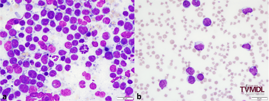
The CBC showed a marked lymphocytosis, marked anemia, and thrombocytopenia (see Table 1). A pathologist reviewed the blood smear and lymph node aspirates. The lymph node aspirates showed a monomorphic population of intermediate to large lymphocytes (see Figure 1a). Similar cells were observed in the blood smear (see Figure 1b). A diagnosis of juvenile lymphoma was made.
| Result | Flag | Reference Interval | Unit | |
| Total WBC | 55.90 | H | 4.0 – 12.0 | K/µL |
| Neutrophils | 0.56 | L | 0.6 – 5.4 | K/µL |
| Lymphocytes | 55.34 | H | 1.8 – 9.0 | K/µL |
| RBC | 2.53 | L | 5.0 – 10 | M/µL |
| HGB | 3.4 | L | 8.0 – 15.0 | g/dL |
| HCT | 10.2 | L | 24.0 – 46.0 | % |
| Platelets | 44 | L | 100 – 800 | K/µL |
| Fibrinogen | 400 | 300 – 700 | mg/dL |
Table 1: Select CBC data from a 5-month-old calf with juvenile lymphoma.
Juvenile or calf lymphoma is a sporadic form of bovine lymphoma that typically presents in cattle younger than 6 months of age. This lymphoma is considered spontaneous and is not associated with enzootic bovine leukosis (EBL) or infection with bovine leukemia virus (BLV). The prevalence of juvenile or calf lymphoma in United States cattle has not been reported but it is considered a rare form of bovine lymphoma.
The most common age of onset of clinical disease ranges from 3 to 6 months. Patients typically present with a history of weakness, weight loss, and moderate depression despite a good appetite. Clinical signs can occur over time or can have an acute onset. Younger calves may be presented for generalized lymphadenopathy. Other clinical abnormalities may include, but are not limited to, cough, anemia, respiratory distress, bloat, and diarrhea.
Typical physical exam findings include generalized bilateral lymph node enlargement, pale mucous membranes, tachycardia, tachypnea, and harsh respiratory sounds on auscultation. Other reported physical exam abnormalities include fever, hyperpnea, ruminal tympany, liver enlargement, and ataxia. Rare cases present with regionalized lymph node enlargement, rather than generalized. In either case the lymph nodes are enlarged but not hot or painful.
CBC findings include a marked lymphocytosis, severe anemia, and possible thrombocytopenia and neutropenia. The anemia, thrombocytopenia, and neutropenia occur secondary to myelopthesis, or bone marrow crowding by neoplastic cells. Affected calves tend to have low serum globulins and elevated aspartate aminotransferase (AST).
The clinical disease syndrome is rapidly progressive and calves typically die within 2 to 8 weeks of the onset of clinical signs. Postmortem histopathology may reveal neoplastic involvement of a variety of organs, especially if there are neurologic abnormalities before death. Neoplastic tissue is found only as small, microscopic nodules.
The etiology of juvenile lymphoma is unknown. The disease is considered to be noninfectious and not contagious. In most cases only individual animals within a herd are affected.

Figure 1: 5-month-old calf with juvenile lymphoma. a) Lymph node aspirates showed a monomorphic population of intermediate to large lymphocytes with some lysed cells and mitotic figures. B) Blood smear examination showed a similar lymphoid population with increased white space between RBCs (anemia) and a thrombocytopenia.