Equine asthma diagnosed with BAL Cytology
Julie Piccione, DVM, MS, DACVP and Judith Akins, DVM, MS
A 23-year-old American Quarter Horse mare was presented to a veterinarian for chronic coughing and exercise intolerance. On physical examination, wheezing was heard during auscultation. Complete Blood Count (CBC) and chemistry profiles were unremarkable. Respiratory endoscopy revealed mildly increased airway mucus. A bronchoalveolar lavage (BAL) was performed and fluid was submitted to TVMDL for cytologic evaluation. A thorough clinical history was provided with the submission.
The BAL fluid was milky and flocculent. Direct, concentrated, and cytocentrifuge slides were made for cytologic evaluation. The slides were highly cellular and non-hemodiluted with occasional dense aggregates of amorphous to fibrillar mucus. Nucleated cells were almost exclusively non-degenerate neutrophils with fewer alveolar macrophages and rare uniform respiratory epithelial cells (Figure 1). No infectious agents were observed cytologically. Given the clinical history and the cytologic findings, a diagnosis of severe equine asthma was made.
Equine asthma is the broadly accepted term used to cover non-infectious, inflammatory, lower airway diseases in horses. The nomenclature for these inflammatory processes has changed over the years. Most recently, mild to moderate equine asthma was previously termed Inflammatory Airway Disease (IAD) and severe equine asthma was termed Recurrent Airway Obstruction (RAO). The pathophysiology of equine asthma is unclear; however, environmental debris and non-infectious agents are presumed to induce hyper-responsive airways and inflammation.
As seen in this patient, severe asthma is most common in older horses. Clinical signs typically include exercise intolerance, coughing, weight loss, and increased respiratory effort at rest. Affected horses often have increased allergen exposure (stables or certain pastures) and may have a genetic history of equine asthma. Cytologic examination of bronchoalveolar lavage fluid reveals marked neutrophilic inflammation, often without substantial bacterial components. Providing clinical history with cytologic submissions improves the odds of achieving a cytologic diagnosis. Severe equine asthma is considered a recurrent and progressive disease; however, clinical signs can be controlled with appropriate therapy.
For more information on clinical pathology testing, visit tvmdl.tamu.edu.
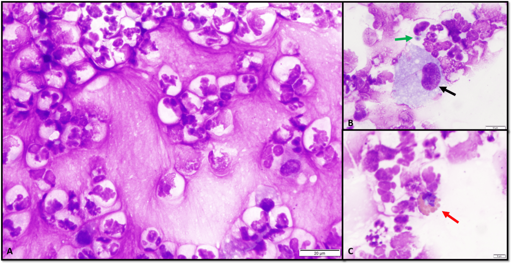
Figure 1: Cytocentrifuge preparation of BAL fluid from a 23-year-old mare. A) Many non-degenerate to poorly preserved (stored sample) neutrophils in a dense mucinous background. B) Macrophage (black arrow) and neutrophil with phagocytized environmental debris (green arrow). C) Eosinophil (red arrow).