Carcinomatosis in laying chickens
Martin Ficken, DVM, PhD, ACVP
A female Gray Barred chicken was presented to the Texas A&M Veterinary Medical Diagnostic Laboratory (TVMDL) in Gonzales. The chicken was approximately 2 years old and weighed 1.41 kilograms.
Following necropsy evaluation, it was noted that within the coelomic cavity there were numerous 1-4 mm in diameter white nodules on the serosal surface of the ventriculus, pancreas, and intestines and on the surface of the mesentery (Figure 1). A 10-cm segment of the anterior oviduct was mildly dilated and covered with confluent nodules similar to those described previously (Figure 2). The ovary was regressing.
Feed was present in both the crop and gizzard. No lesions were noted in any other tissue or organ system. The diagnosis was determined to be carcinomatosis.
In a similar case, one dead adult female chicken was presented for necropsy to the Gonzales laboratory. History noted the chicken was found dead on the coop floor following a decrease in appetite over the previous two to four days.
Upon necropsy evaluation, the bird had moderate breast muscle atrophy and weighed 1.83 kilograms. Approximately 200 ml of straw-colored fluid was present in the coelomic cavity. The liver contained multiple polycystic nodules throughout the parenchyma (approximately 2 mm to 2 cm in diameter). On the ventral surface of the left liver lobe there was a large multicystic mass adhered to the capsule.
The spleen had multiple 1 mm nodules on the capsular surface. The intestinal loops were adhered to one another with the serosal surfaces and omentum covered with numerous small white nodules.
The ovary had numerous white nodules that were approximately 1-mm. The oviduct had multiple 1 to 5 mm nodules on the serosal surface. The right oviduct was persistent and has two cysts approximately 3 cm in diameter.
The lungs were severely edematous and congested. Following necropsy, diagnoses were determined to be ovarian carcinoma, presumptive, with carcinomatosis and ascites.
Carcinomatosis, within the coelomic cavity of chickens, is a common neoplastic condition in adult female chickens. The condition is due to metastatic adenocarcinomas arising from the ovary, oviduct, or pancreas. In many instances, as observed in the second case, ascites is present in the coelomic cavity. Histopathology is often unrewarding in determining the tissue of origin. These tumors are considered spontaneous and are not related to any known tumor-inducing avian virus (Marek’s, avian leukosis, reticuloendotheliosis, other avian retroviruses).
Reference
- Hafner S, Reece RL, Williams SM. Neoplastic Diseases (Other Tumors) in Diseases of Poultry, 13th edition, Wiley-Blackwell, ed. Swayne, DE et al. pp. 605-608, 2013.
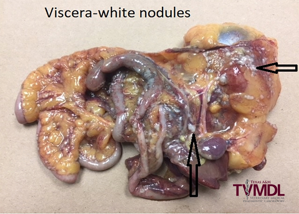
Figure 1 – Numerous 1-4 mm in diameter white nodules on the serosal surface of the ventriculus, pancreas, and intestines and on the surface of the mesentery.
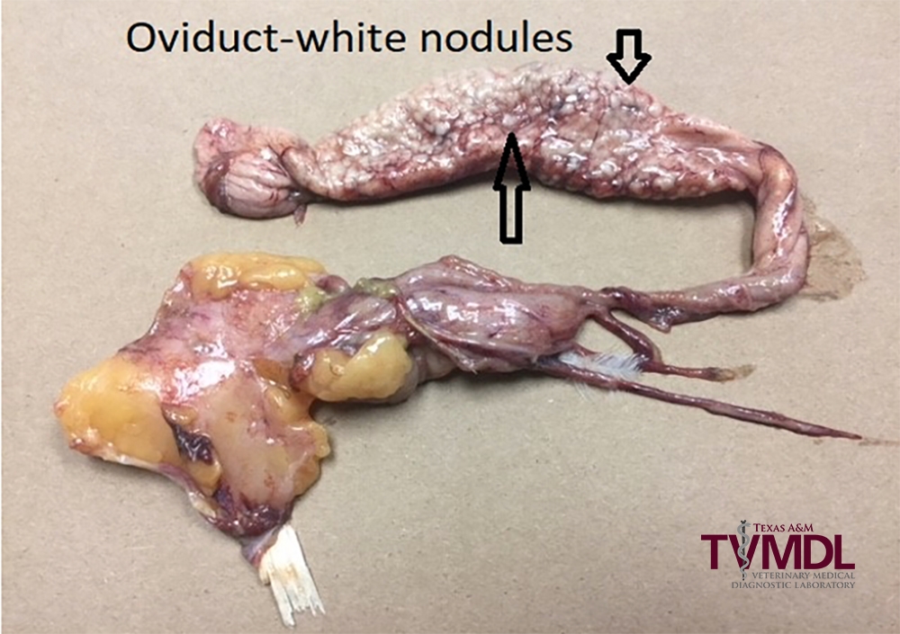
Figure 2 – Oviduct covered with coalescing neoplastic nodules.