Osteogenesis and Dentinogenesis Imperfecta in a Calf
Randi Gold VMD, PhD, DACVP and Guy Sheppard DVM
A 21 day-old calf with a history of multiple leg fractures and no evidence of external injuries was euthanized and sent into the TVMDL for evaluation of the hind limbs and mandible. On gross necropsy examination the mandible contained incisor teeth that were erupted but were all pink, translucent, mobile, and when transected contained pink gelatinous material (Figure 1). Both hind legs contained tibial fractures. The right was acute and oblique with marked surrounding hemorrhage and edema. The left was chronic with mild callus formation. Cortices of the leg bones were thin along the length of the shafts.
Histopathology of the left cannon bone demonstrated diffuse osteopenia with decreased to absent cortices. The trabecular bone was markedly thin, irregular, basophilic, and had maintenance of the cartilage cores as the trabeculae transitioned from the primary to secondary spongiosa (Figure 2). Osteoblasts varied from plump to thin and attenuated. There were multifocal microfractures.
Within the incisor teeth, the dentin was irregular with disorganization of the dentin tubules (dysplasia) (Figure 3). Odontoblasts were irregular, disorganized and had a loss of polarity. There was variable thickness of the predentin. Multifocally ameloblasts were shrunken, cuboidal to polygonal, and disorganized with increased cell layer piling (dysplasia). The underlying and supporting alveolar bone was osteopenic.
Osteogenesis imperfecta is an inherited connective tissue disorder characterized by excessive bone fragility with a wide array of clinical signs and variations in severity. The majority of mutations are either in the COL1A1or COL1A2 genes, which code for type 1 collagen. Abnormal clinical findings can include spontaneous fractures, fragile, iridescent teeth (dentinogenesis imperfecta), joint laxity, and blue sclera. The primary differential diagnoses are nutritional/metabolic bone disease and trauma, most of which carry a more favorable long term prognosis if corrected. Osteogenesis imperfecta in cattle and small ruminants has previously been documented in the Angus, Hereford, Charolais, Belgian blue, and Holstein-Friesian cattle breeds and Romney sheep. In dogs it has been seen in several breeds including golden retrievers, beagles, collies, poodles, Norwegian elkhounds, Bedlington terriers, dachshunds, a Chow Chow, and recently a Mastiff. Unfortunately, medical management is often unsuccessful.
References
– Craig LE, Dittmer KE, Thompson KG. Bone and Joints. In: Maxie MG, editor. Jubb, Kennedy, and Palmer’s Pathology of Domestic Animals. 1. 6 ed. Missouri: Elsevier; 2016:17-163.
– Gold R, Pool RR, Edwards EE. Osteogenesis and dentinogenesis imperfecta in a four-month-old English mastiff. Vet Rec Case Reports 2019;7: e000835. doi: 10.1136/vetreccr-2019-000835
– Thompson KG. Skeletal diseases of sheep. Small Ruminant Res 2008;76:112-9.
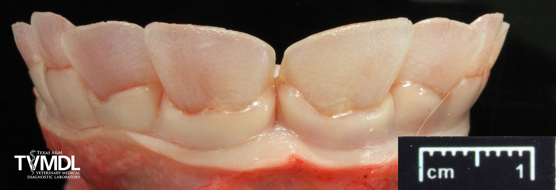
Figure 1: Mandibular incisor teeth. The teeth are erupted but are all pink, translucent, and easily breakable.
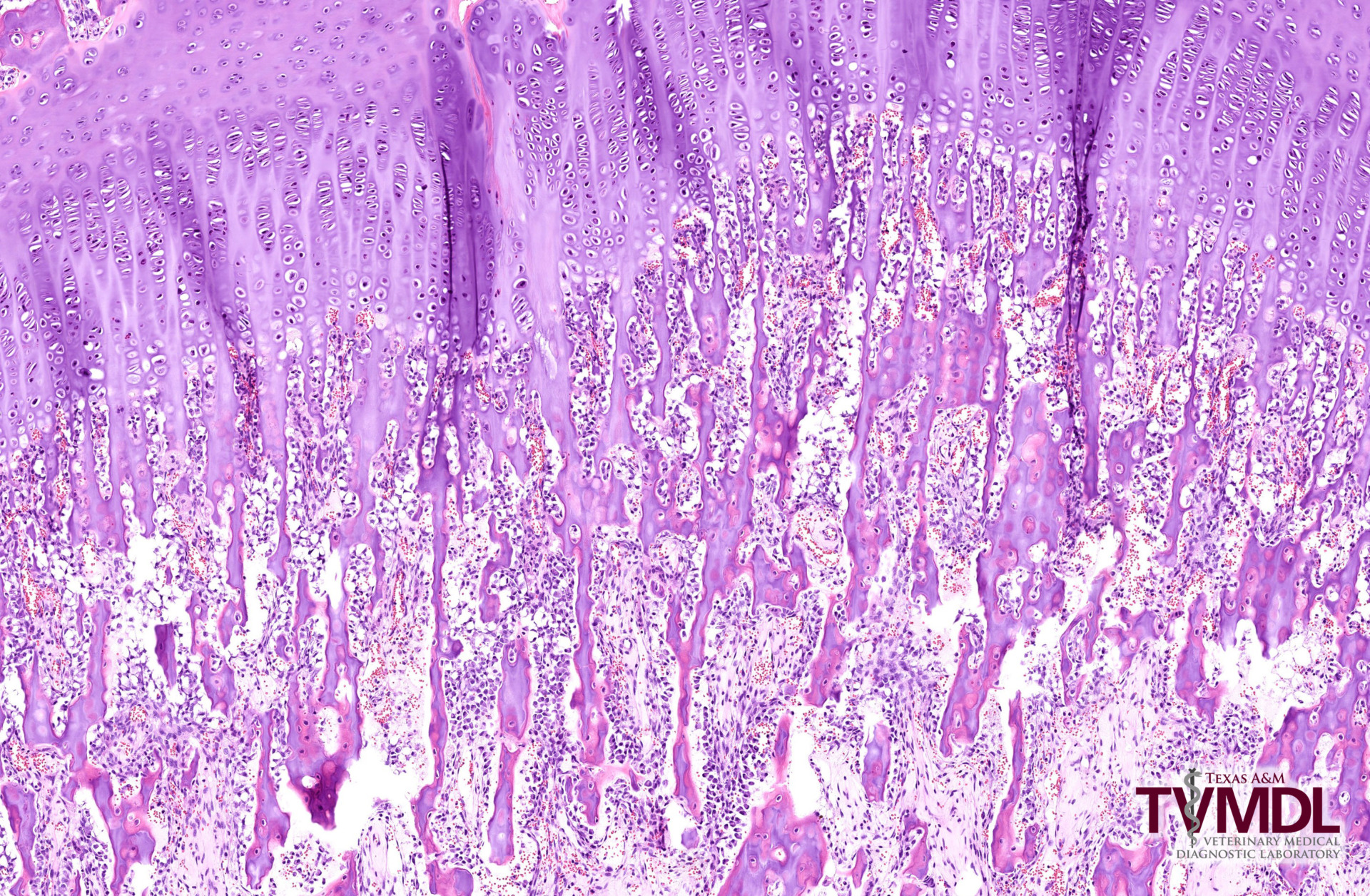
Figure 2: H&E of the epiphysis of the left cannon bone. There is maintenance of the cartilage cores as the trabeculae transitioned from the primary to secondary spongiosa.
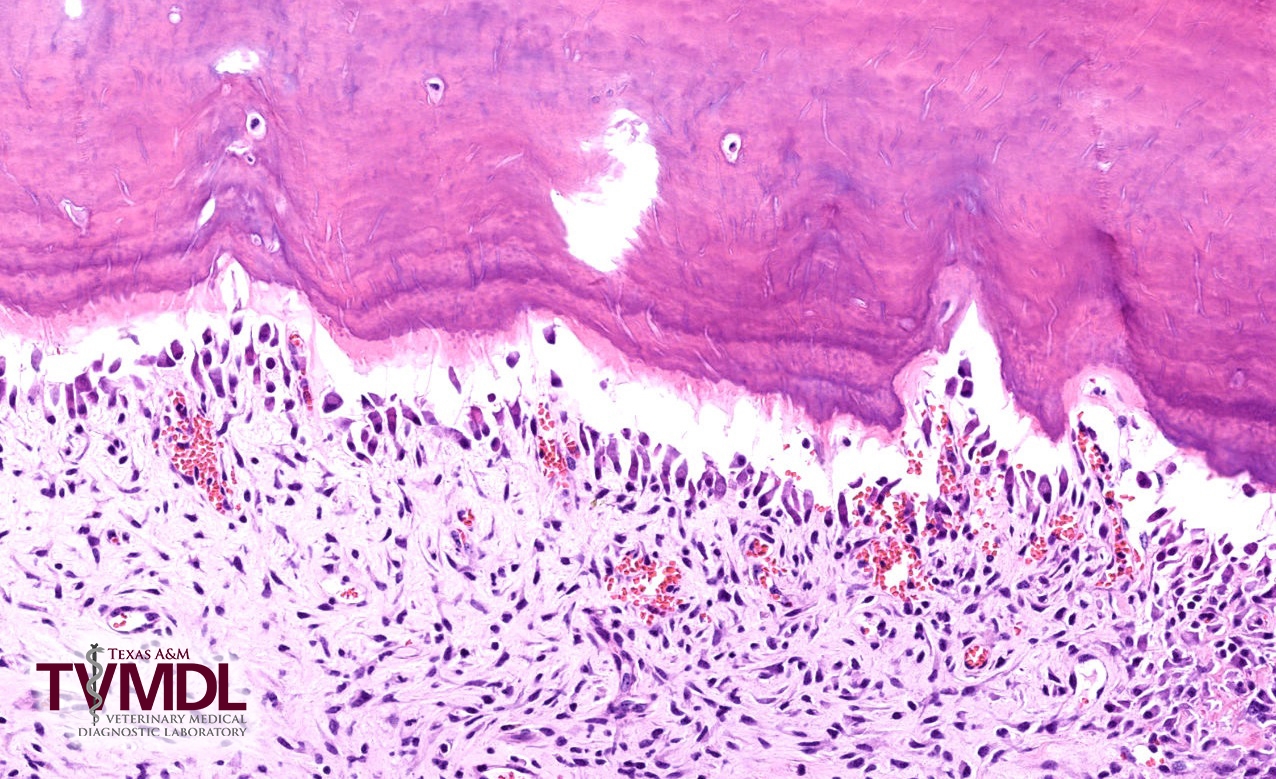
Figure 3: H&E of an incisor tooth. There is irregularity and disorganization of the predentin and dentin. Odontoblasts are disorganized and have decreased polarity.