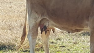
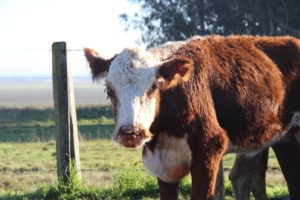
Photosensitization is a serious skin condition in horses and cattle caused by a hazardous combination of certain plants and ultraviolet (UV) light. Certain plants contain photodynamic agents, which then cause a reaction in the animal’s body that leads to ultra-sensitive skin. This condition is specific to lightly or non- pigmented animals or areas of an animal that have less hair. Severe skin damage can result and include ulceration, necrosis, and edema.
Photosensitization is generally classified by the source of the photodynamic agent. The three typical classifications are:
- Primary Photosensitization (Type I): Occurs when a photodynamic agent is injected, ingested, or absorbed in the skin. Once an agent enters the systemic circulation and is exposed to UV light, damage to the skin’s cell membrane occurs.
- Aberrant Pigment Metabolism (Type II): Syndrome in which autogenous pigments are photosensitizing porphyrin agents. This syndrome is a congenital defect. (uroporphyrin I, coproporphyrin I, protoporphyrin III)
- Secondary (Hepatogenous) Photosensitization (Type III): Most common form observed in livestock. Occurs when an animal ingests plants that contain phylloerythrin. If the animal has liver damage, phylloerythrin will not be excreted into the animal’s bile and will eventually be circulated. When phylloerythrin reaches the skin, it will initiate a photoxic reaction, causing severe skin burns and sloughing.
Causative Toxic Plants
Primary photosensitization:
- Bishop’s weed (Ammi majus) furocoumarin
- Rainlily (Cooperia pedunculata) fungal elaboration
- Dutchman’s breeches (Thamnosma texana) psoralens
- St. John’s Wort (Hypericin)
- Buckwheat (Fagopyrin)
- Coal tar derivatives such as tetracyclines and polycyclic aromatic hydrocarbons
Hepatogenous photosensitization :
- Moldy bermudagrass (Cynodon dactylon) fungal elaboration
- Lantana (Lantana camara) lantadene A and B
- Sacahuista, blooms only (Nolina texana) saponins
- Kleingrass (Panicum coloratum) saponins
- Puncturevine (Tribulus terrestris) saponins
Clinical Findings and Lesions
Regardless of the cause, classical signs of photosensitization are similar. Classical signs of a photosensitive animal are:
- Immediate discomfort and agitation when exposed to light
- Rubbing or scratching of lightly pigmented or exposed skin areas
- Lesions in exposed areas
Main lesions include skin necrosis and ulceration of exposed areas. Exposed areas include, but are not limited to:
- Face
- Sides of udders
- Underside of tongue
- Lips
- Eyelids
- Ears
Differences in distribution are related to differences in skin pigmentation of breeds and individual animals Affected skin will be red, oozing fluid, and swollen. In severe cases, large crusts of necrotic, black skin
may slough off. Affected animals may also be icteric with yellow discoloration of the skin and mucus membranes. Animals may also experience blindness due to corneal opacity.
Testing Recommendations
TVMDL recommends submitting samples for the following tests. In the case of photosensitization, rumen microscopy is of limited use.
To download and print this information, visit the Bovine section of the Education Library.
For more information on TVMDL’s testing services, visit tvmdl.tamu.edu or call one of the agency’s four laboratories.

