Cutaneous Pythiosis in a German Shepherd Dog
Cheryl Maguire, DVM, Dominique Wiener, DVM, PhD, DECVP, Sonia Lingsweiler, BS, MS
The bacteriology section at the Texas A&M Veterinary Medical Diagnostic Laboratory (TVMDL) received a skin swab for bacterial culture and susceptibility from a 1-year old, female, German Shepherd dog with a history of a non-healing wound over the caudal dorsum (Figure 1) that was suspected to have resulted from a dog bite. Multiple organisms including Staphylococcus intermedius Group were isolated, all of which were susceptible to all antibiotics tested aside from Ampicillin.
The wound did not respond to therapy and at the time of follow-up to the referring veterinarian the dog presented with a fever (104.6F), and the lesion had spread and contained multiple draining tracts and pustules. After consultation with a TVMDL veterinary diagnostician, the referring veterinarian submitted multiple biopsies fixed in formalin for dermatopathology as well as fresh tissue for bacterial and fungal cultures.
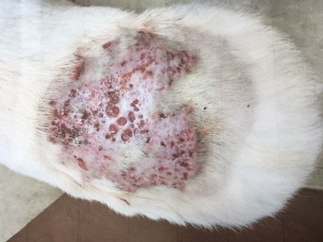
Figure 1. Non-healing wound over the caudal dorsum.
Dermatopathology Results
In the deep dermis and subcutis, there were multifocal to coalescing, many epithelioid macrophages, multinucleated giant cells, and neutrophils that were multifocally forming pyogranulomas (Figure 2 A and B). In the periphery of the granulomas, there was a rim of lymphocytes, plasma cells, and proliferating fibroblasts (fibrosis). Multifocally, there were free erythrocytes intermixed with the lesions (hemorrhage). In the center of the granulomas and within multinucleated giant cells, there were negatively-staining fungal hyphae (Figure 2C). In the GMS stain the fungal hyphae were 6-12μm wide, had non-parallel walls and are were poorly septate (Figure 2D). A morphologic diagnosis of a severe, chronic, multifocal-coalescing pyogranulomatous dermatitis and panniculitis with intralesional fungal organisms was made.

Figure 2: Skin biopsy (dermis and panniculus), dog. A) In the deep dermis and panniculus, there is a multifocal-coalescing severe inflammation; scale bar= 500μm. H&E stain; 2x magnification. B) Many epithelioid macrophages, multinucleated giant cells, and neutrophils are forming pyogranulomas. In the periphery are lymphocytes and plasma cells. H&E stain; 10x magnification; scale bar= 100μm. C) Epitheloid macrophages and multinucleated giant cells surround or engulf negatively-stained fungal hyphae (black arrows). 60x magnification; scale bar= 20μm. D) Fungal hyphae are 6-12μm wide, have non-parallel walls and are poorly septated. GMS stain; 20x magnification; scale bar= 50μm. Inset: Higher magnification of D). GMS stain; 60x magnification; scale bar= 20μm.
Microbiology Results
Upon arrival, the fresh tissue biopsy was macerated and plated on routine bacterial and fungal culture media. After overnight incubation, a swarming fungus-like growth was present which was morphologically compatible with Pythium. Because we cannot differentiate between Pythium and Lagenidium species (closely related organisms in the class Oomycetes), the isolate was forwarded to the molecular lab for sequencing and the identification was confirmed to be Pythium insidiosum.
Case Summary
Pythium insidiosum is an aquatic pathogen that uncommonly causes disease of the gastrointestinal tract or skin of dogs and more rarely the skin of cats. Young, large-breed dogs that have access to wet environments such as ponds, lakes and bayous are more likely to become infected. German Shepherd Dogs may be predisposed to the cutaneous form. Horses can also develop pythiosis.
In severe, chronic, and/or non-responsive dermatological cases, biopsy for dermatopathology with concurrent tissue culture is recommended. To learn more about these services or how to collect and submit samples, please visit tvmdl.tamu.edu or call 1.888.646.5623.
Reference:
Grooters AM. Pythiosis, Lagenidiosis, and Zygomycosis. In: Sykes JE ed. Canine and Feline Infectious Diseases. St. Louis, MO: Elsevier Saunders, 2014.