A tale of a tail tumor: Ferret chordoma
Erin Edwards, DVM, MS, DACVP
Chordomas are among the most common diagnoses in ferret biopsy samples submitted to the Texas A&M Veterinary Medical Diagnostic Laboratory (TVMDL). This article highlights a recent case of a ferret chordoma that was diagnosed at our laboratory. The tumor was located at the tip of the tail of a 3-year-old ferret. The tail tip was amputated and sent to TVMDL for biopsy, and histopathologic examination showed classical features of a chordoma.
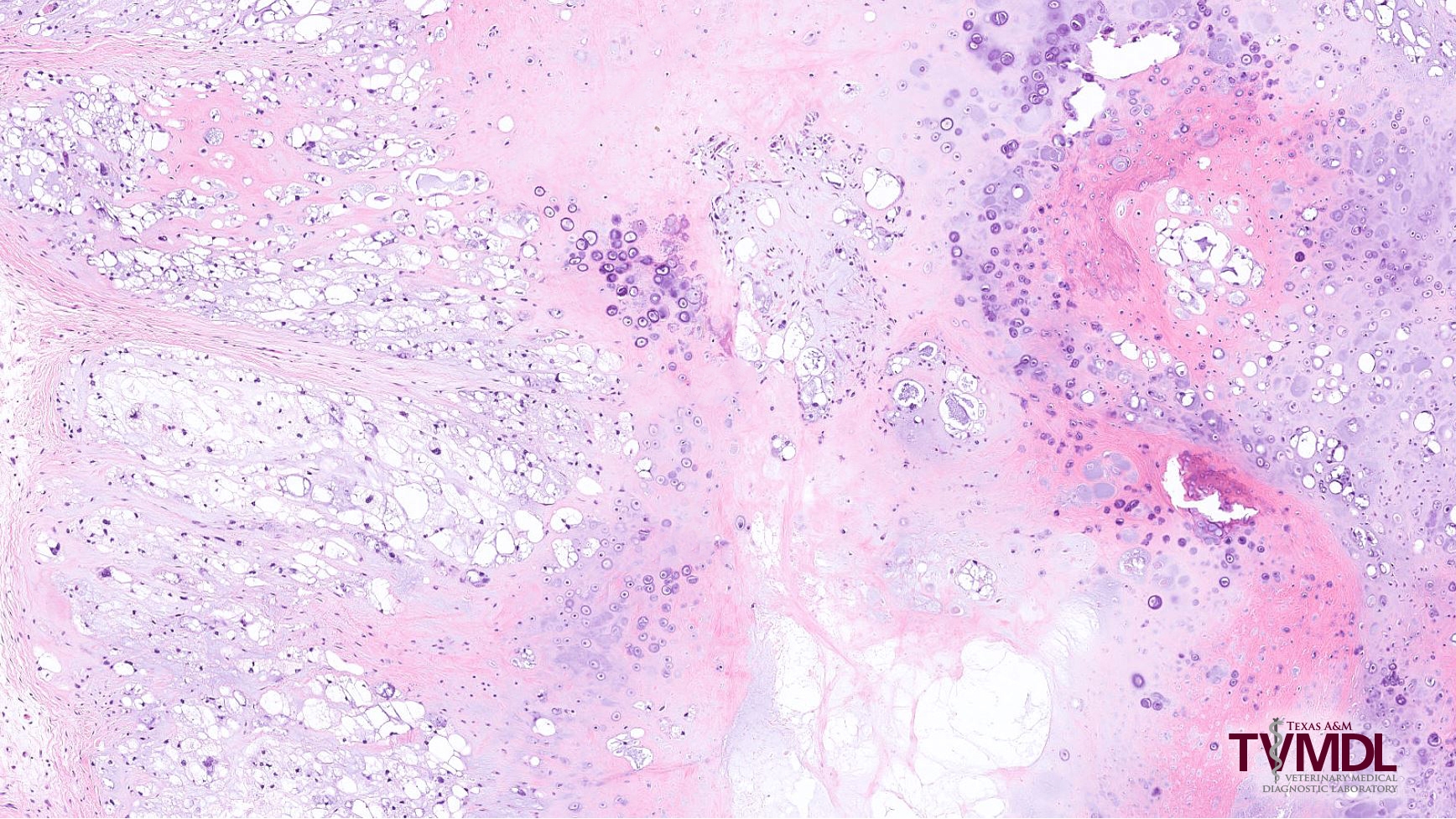
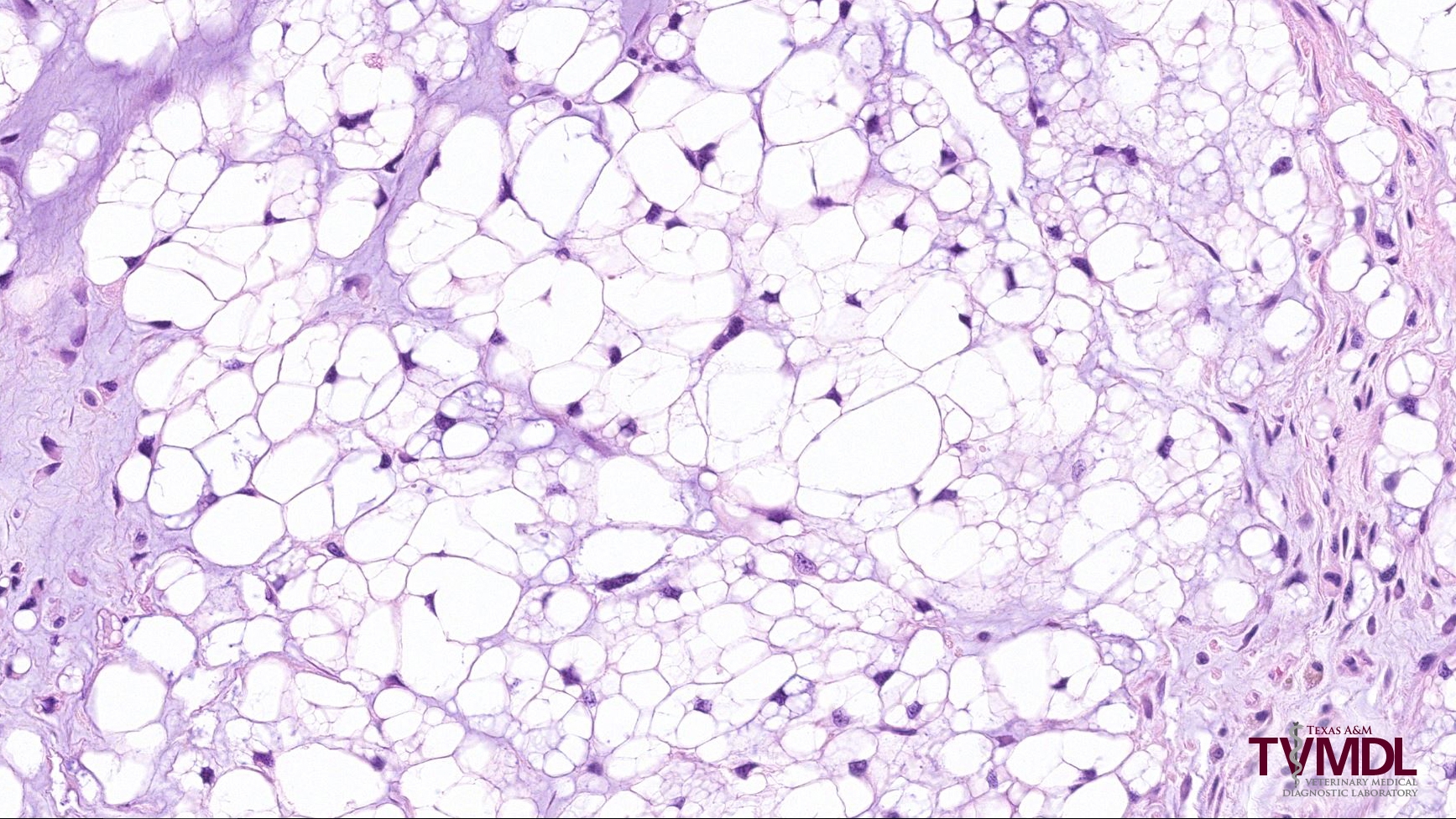
Chordomas are common tumors of ferrets and are rarely diagnosed in other species. They are thought to develop from intraosseous remnants of the fetal notochord. In ferrets, these tumors tend to occur on the tail, usually at the tail tip. They can less commonly occur in the cervical spinal column or rarely along other sites of the spinal column. Histologically, chordomas variably contain bone, cartilage, and characteristic physaliferous cells (Figure 1). Physaliferous cells have a vacuolated, foamy cytoplasm that is often compared to soap bubbles (Figure 2).
Chordomas are typically slow-growing masses. They can be locally invasive and destructive, leading to local discomfort with possible altered ambulation or, in severe cases, paralysis. Fortunately metastasis is rare. Chordomas occurring at the tip of the tail can often be cured with surgical excision.
References:
– Fox, J. G., Muthupalani, S., Kiupel, M., & Williams, B. (2014). Neoplastic diseases. In J. G. Fox & R. P. Marini (Eds.), Biology and Diseases of the Ferret (3rd ed., pp. 612-613). Ames, IA: John Wiley & Sons.
– Schoemaker, N. J. (2017). Ferret oncology: diseases, diagnostics, and therapeutics. Veterinary Clinics: Exotic Animal Practice, 20(1), 183-208.

