• Skip to primary navigation
  • Skip to main content
  • Skip to primary sidebar
  • Skip to footer
  • Locations:
  • Canyon
  • Center
  • College Station
  • Gonzales
  • TVMDL Career Center
  • Contact Us

Texas A&M Veterinary Medical Diagnostic Laboratory

Apple App

download on the Apple App Store
download the app on Google Play

Search

Translate:

  • Find a Form
  • Deliver a Sample
    • Specimen Collection Information
    • Packaging Samples for Shipment
    • Drop Off a Sample
    • Cremation
    • Order Supplies
  • Client Portal
    • Get Test Results
    • SVA Portal
    • Pay a Bill
  • Become a Client
  • Resources
    • TVMDL Bovine Testing Guidance and Reference Material
    • Diagnostic Plans
      • Bovine Syndromic Diagnostic Plans
      • Equine Syndromic Diagnostic Plans
      • Small Ruminant Syndromic Diagnostic Plans
      • Small Animal Syndromic Diagnostic Plans
    • Education Library
    • Case Study Library
    • Electronic Reporting: QR Coding Process
    • CoreOne Resources
  • About Us
    • Locations and Hours
    • Agency Leadership
    • Speaker’s Bureau
    • Texas Pullorum-Typhoid Program
    • TVMDL Mobile app
    • Contact Us
  • Locations
    • College Station Laboratory
    • Canyon Laboratory
    • Center Laboratory
    • Gonzales Laboratory
  • Contact Us

Fungal Equine Endometritis

May 5, 2021 by Mallory Pfeifer

Fungal Equine Endometritis
Judith Akins, DVM, MS

Vaginal/uterine discharge was submitted to the Texas A&M Veterinary Medical Diagnostic Laboratory (TVMDL) in College Station from an eight-year-old Warmblood mare for culture and a cytologic evaluation. Cytologically the discharge consisted of large numbers of degenerate neutrophils and marked numbers of yeast. The neutrophils were actively phagocytizing the yeast. There were no bacteria identified. The culture yielded a 4+ growth of yeast. Fungal endometritis was diagnosed.

Fungal endometritis in the mare is less common than bacterial infections. The more common sources for a fungal infection are the skin adjacent to the vagina and fecal contamination. The most commonly isolated fungi include Candida spp. and Aspergillus spp. Pneumovagina, chronic endometritis, and prolonged use of intra-uterine antibiotics may all play a role in allowing fungal yeast or hyphae to overgrow and cause disease.

For more information on TVMDL’s bacteriology or clinical pathology test offerings, visit tvmdl.tamu.edu or call the agency’s College Station laboratory.

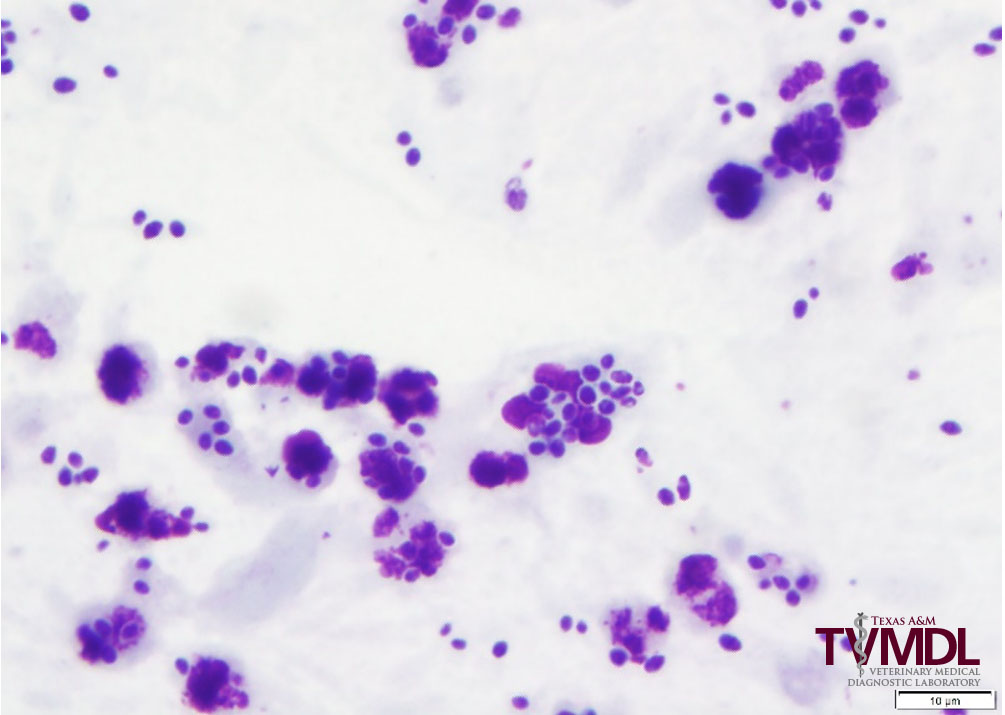
Figure 1. Yeast and neutrophils in uterine fluid. 100x objective.

Filed Under: Case Study Tagged With: clinical pathology, equine, horse, pathology

Primary Sidebar

Latest Case Studies

  • Neurologic Disease due to Bovine Herpesvirus-5 (BHV-5) Infection in a 3-week-old Charolais mix calf

    February 27, 2023

  • Rare case of ocular onchocerciasis in a dog from south Texas

    January 25, 2023

  • Cerebrospinal Nematodiasis (Visceral Larval Migrans) in Birds

    September 13, 2022

  • Spironucleosis (Hexamitiasis) in Quail

    August 9, 2022

  • Endocarditis in a White-Tailed Deer Caused by Trueperella pyogenes

    August 9, 2022

Footer

For Employees

  • Employee Email
  • TVMDL Rules & Procedures
  • TVMDL Career Center
  • Emergency Alert Systems
  • AgriLife People Directory
  • AgriLife Administrative Services
  • TAMUS Single Sign On
  • Facebook

State of Texas

  • Texas.gov Portal
  • Texas Veterans Portal
  • Statewide Search
  • Texas Homeland Security
  • Risk, Fraud, & Misconduct Hotline

Policies

  • Privacy and Security Policy
  • Accessibility Policy
  • Texas A&M AgriLife
  • Texas A&M Veterinary Medical Diagnostics Laboratory
  • Texas A&M Forest Service
  • Texas A&M AgriLife Extension Service
  • Texas A&M AgriLife Research

483 Agronomy Rd
College Station, TX 77843