Submitting samples of bone for histopathological analysis is essential to definitively diagnose most primary bone diseases (e.g., osteosarcoma). However, collecting representative bone biopsy specimens and their histopathological interpretations presents several challenges for the clinician and the pathologist, respectively.
Tips for Collecting Needle and Core Bone Biopsy
The majority of osteosarcomas are central/medullary (i.e., the tumor starts off within the medullary cavity, grows until it fills this space, and then breaches the outer cortex of the bone). Most of the time the surface just contains reactive bone with a proliferative periosteal surface. If a needle or trephine biopsy sample only represents the surface of the bone and does not breach the cortex and reach the endosteum, there is a good chance that the histopathologic diagnosis will reflect these non-specific changes and not give the true diagnosis of osteosarcoma. When possible, samples should be collected from deeper within the lesion as they are more likely to be diagnostic. We encourage you to send relevant diagnostic images and/or imaging reports (i.e. radiographs, CT, MRI images) with your submission. These can be emailed to clientservices@tvmdl.tamu.edu and can aid in pathologic evaluation and interpretation. Tips to help you improve the chances of getting a diagnosis when submitting a needle or core bone biopsy include:
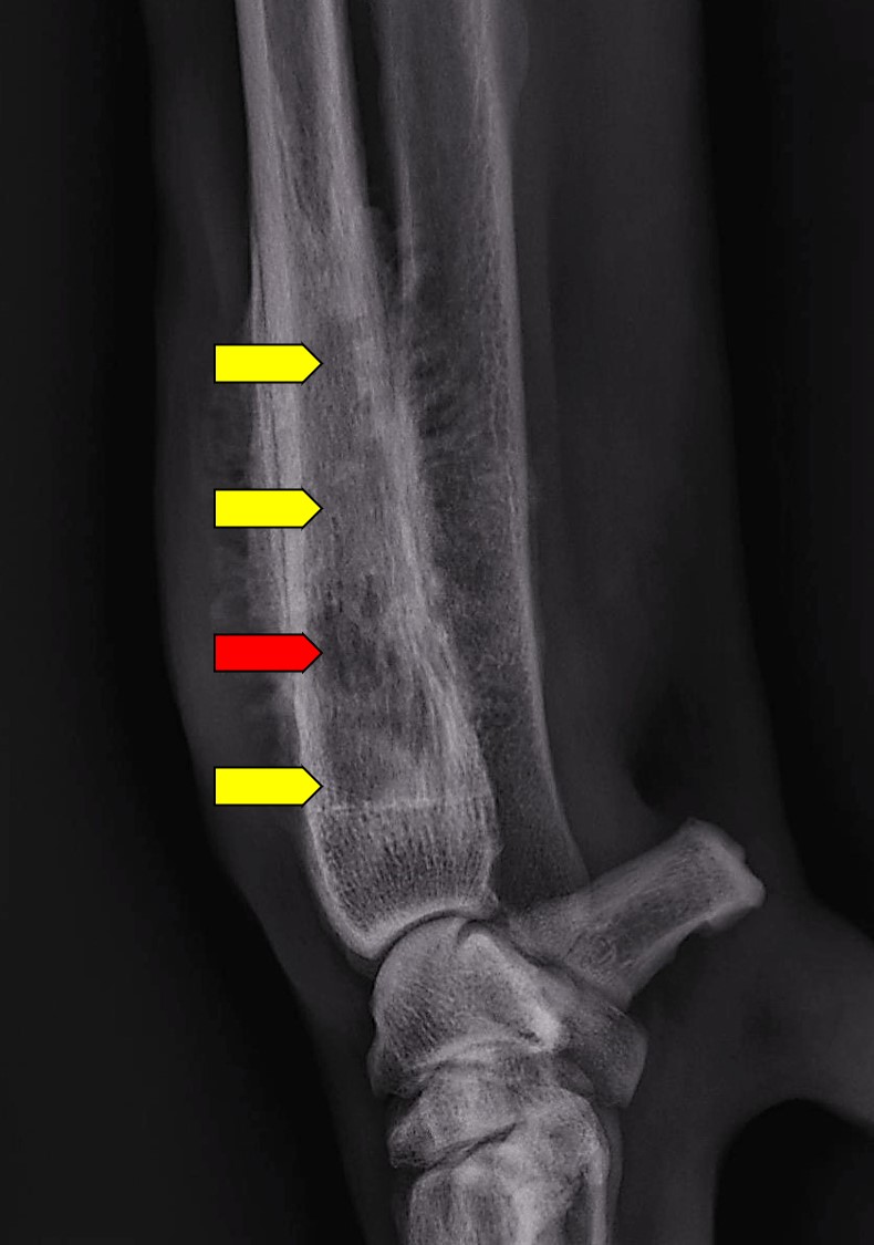
Figure 1. Radiograph of an osteosarcoma on the distal radius. Red arrow shows the center of the mass which may be necrotic due to devitalization. Yellow arrows depict areas of sclerosis and transition between normal and abnormal bone. Recommend all sites be sampled for thorough osteopathic evaluation.
1. Minimizing artifact
During the procedure, tissue handling should involve a balance between the force needed to collect an adequate sample with delicate handling to avoid crush/squeezing artifacts that compromise the microscopic characteristics of the biopsy. For example, using care while transferring the biopsy from the tip of the needle into formalin is a stage this can be accomplished. Most samples should be promptly placed in 10% neutral buffered formalin once collected.
2. Take multiple sections
All bone needle biopsy specimens are a relatively small portion of the whole bone that may not be representative of the entire bone lesion. Therefore, collecting multiple samples will maximize your chances of submitting representative and diagnostic tissue.
3. Sample different areas of the lesion (Figure 1)
Taking a sample from only the center of the lesion may yield negative diagnostic results, as the center of the tumor is often the first location to become devitalized due to a lack of blood supply (red arrow). The center should be still be sampled, but you should also sample areas of sclerosis or areas of transition between normal and abnormal sections of bone for a better likelihood of getting diagnostic tissue (yellow arrows).
4. Save a fresh sample or swab
If you are considering bacterial or fungal disease, swab the affected site or keep a fresh sample for culture before placing the entire sample in formalin. These samples can be submitted up front for testing or held pending histologic evaluation.
Tips for submitting an entire leg
Fresh or formalin-fixed whole legs can be submitted to TVMDL. Additional fees apply when submitting either as these require additional sectioning, processing, and gross examination by a pathologist. There are pros and cons of each (see below). In these situations, radiographs are often extremely helpful for lesion localization and limb dissection, especially when there are no obvious lesions on gross examination of a whole limb. These can be emailed to clientservices@tvmdl.tamu.edu.
Submitting in formalin
- One advantage of submitting limbs in formalin is that fixation will start immediately, reducing the length of time needed for tissue processing and overall report turnaround time.
- If sending a limb in formalin, do not wrap it in tape or vet wrap as the formalin will not penetrate these materials.
- One disadvantage is that formalin will change the color and texture of tissue. This can make it harder to appreciate subtle lesions during gross dissection. If the lesion is in the soft tissue, tagging it with a suture can help us find it.
- Depending on the size of the animal, it may not be possible to find a container with formalin large enough to fit the leg. If this is the case and you want to know if there is part of the limb that you can discard, please don’t hesitate to call the lab to discuss options.
- Another disadvantage of sending a formalin-fixed leg is that samples cannot be collected for bacterial/fungal culture at the lab if histology indicates the need for this testing.
Submitting fresh
- A potential disadvantage is that fixation is delayed, which could result in minor turnaround time delays.
- One advantage is that with fresh tissue, the pathologist may be better able to evaluate the lesion grossly than with formalin fixed tissue.
- If submitting fresh limbs, please pack them with ice packs to ensure they will get to TVMDL in good condition.
- Submitting fresh limbs allow us to collect fresh tissues or swabs for bacterial/fungal culture if indicated.
Clinical history
Knowledge of the animal age and breed can help generate a list of differential diagnoses and aid in histologic interpretation. The location of the lesion should not only include which bone is affected, but also the specific location (i.e. distal metaphysis, growth plate, epiphysis, mid diaphysis, etc.). Previous pertinent medical history and treatment modalities are other considerations, in addition to incidences of previous trauma to the biopsy site, previous tumors in the patient at the biopsy site, or tumors elsewhere in the body, etc.
| Suboptimal Example | Ideal Example | |
| Signalment | Dog | 8-year-old M(N) Rottweiler |
| Brief Clinical History | Lameness | Fracture at biopsy site 2 years ago, repaired via internal fixation, now lytic lesion at repair site |
| Radiographic Findings | Tibia appears abnormal | Send the radiographs and attach the report or brief description of abnormalities |
| Current/Previous Treatment | Not responding to treatment | Currently receiving steroids or antibiotics were started with no improvement |
| Additional Relevant History | History of masses | Had a mammary carcinoma removed 2 years ago. Had a digital squamous cell carcinoma previously removed from the same leg last year. |
Download the printable version of this article in the TVMDL education library.