Hepatocytoptropic lymphoma in a Great Dane
Morgan Matthews, DVM
Tissues from a 3-year-old, castrated male Great Dane were submitted to the Texas A&M Veterinary Medical Diagnostic Laboratory (TVMDL). On histopathology, the liver, spleen, and kidney were infiltrated by neoplastic lymphocytes. Neoplastic lymphocytes migrated through hepatic sinusoids and uniquely, also infiltrated hepatocytes.
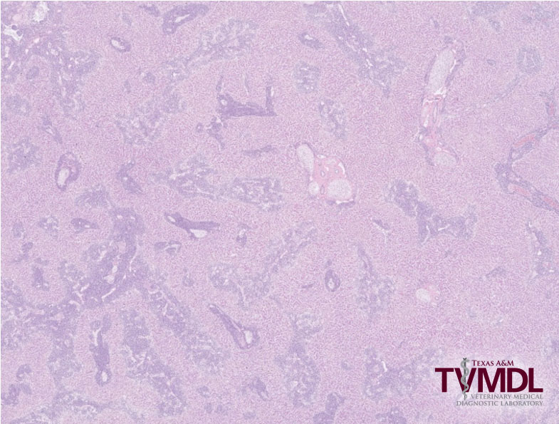
Figure 1. Low power microscopic view of the liver showing effacement of portal areas with neoplastic lymphocytes.
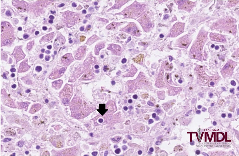
Figure 2: Invasion of hepatocytes by neoplastic lymphocytes.
Hepatocytotropic lymphoma is a rare, highly aggressive form of T-cell lymphoma, arising from a subset of cytotoxic T cells. The neoplasm is rapidly progressive, and dogs often die within 24 hours of presentation. In reported cases, patients presented with thrombocytopenia, hypoalbuminemia, hyperbilirubinemia and cholestasis. Other affected organs may include spleen, lung, and kidney. A related form of lymphoma, arising from the same type of T cell, is hepatosplenic lymphoma and in humans, young to middle aged men are more frequently affected.
For more information on hepatocytotropic lymphoma, contact one of TVMDL’s full service laboratories in College Station or Canyon or visit tvmdl.tamu.edu.
References
Keller, S.W., et al. Hepatosplenic and Hepatocytotropic T-Cell Lymphoma: Two Distinct Types of T-Cell Lymphoma in Dogs. Vet Path. 2012; 50(2): 281-290