Enterotoxemia in Sheep and Goats
Guy Sheppard DVM and Randi Gold VMD, PhD, DACVP
Enterotoxemia, commonly called “overeating disease”, is a common problem in sheep and goats, especially in animals under a year of age. The disease occurs in peracute, acute, and chronic forms. The causative bacterial organism is Clostridium perfringens with types C and D being the most common forms. Type C is said to be seen most commonly in lambs or kids younger than 3 weeks of age (“milk colic”) while Type D is found in older animals. Enterotoxemia is usually seen in rapidly growing feedlot lambs on high concentrate rations, but it is also quite common in fast growing and well-conditioned animals grazing on lush pastures. Animals affected by this condition do not “overeat” as is the case with grain overload. They are simply growing and gaining weight in ideal conditions, and the Clostridium perfringens organism also begins to grow rapidly, releasing its toxins.
Clinical signs of infection are characterized by sudden onset of depression, abdominal pain, diarrhea, neurologic signs, or sudden death. Death usually occurs within hours of the onset of signs. The sudden onset of neurologic signs followed by sudden death is said to be more common in lambs while kid goats are more likely to show signs of diarrhea before death.
Treatment is rarely effective or available, and prevention is far more likely to be successful. Routine vaccination for Clostridium perfringens types C&D in lambs and kids is very effective, but keep in mind that vaccinated animals will require a booster of the product 3 to 4 weeks after the initial vaccination. Given the fact that a related infection known as tetanus is also very common in lambs and kids, producers might want to consider using a combination product than contains tetanus toxoid also.
Should the animal die, a necropsy can be performed to look for evidence of disease. On necropsy examination, a large section of the small intestine can appear dark red to purple (Figure 1). This coincides histologically to necrosis and ulceration of the mucosal surface with diffuse hemorrhage (marked necrohemorrhagic enteritis). The intestinal contents are often bloody and full of fibrin clots and necrotic debris. Petechiation of the epicardium and endocardium may or may not be present.
To confirm the diagnosis, bacterial culture can be performed on a segment of the small intestine and its contents. This can be followed by polymerase chain reaction (PCR) typing to determine the exact strain of the causative bacteria.
For more information on enterotoxemia, contact Veterinary Diagnostician Dr. Guy Sheppard or Veterinary Pathologist Dr. Randi Gold. Learn more about TVMDL’s test offerings, by visiting tvmdl.tamu.edu.
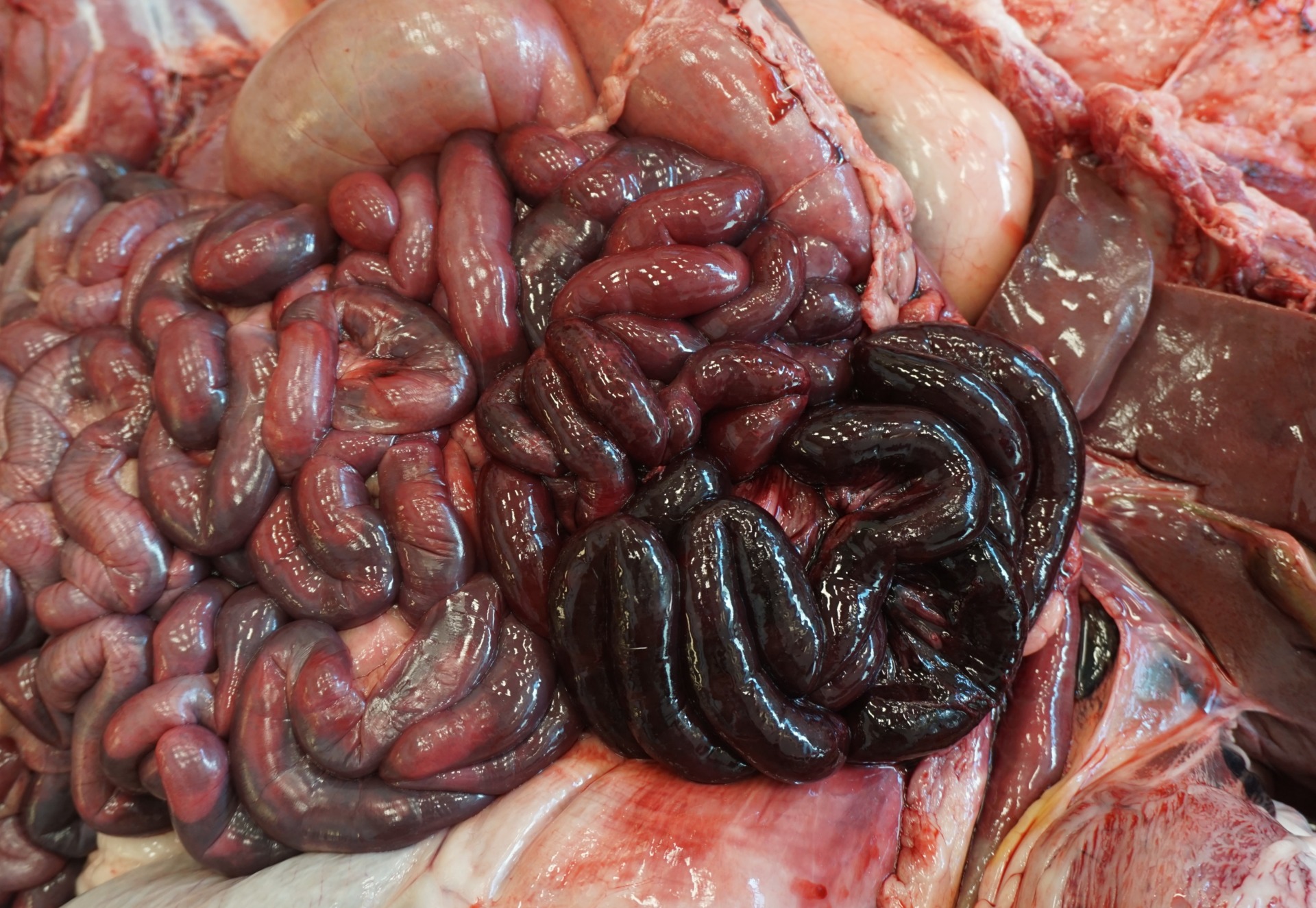
Figure 1: The intestines contain a segment of bowel that is dark red to purple (marked necrohemorrhagic enteritis).
Reference
Smith, M.C., Sherman, D.M., Uzal, F.A., Kelly, W.R., Sheep and Goat Medicine; D.G. Pugh, DVM, editor. 2002 pg. 84.
Uzal, F. A., Plattner, B. L., & Hostetter, J. M. (2016). Alimentary system. In M.G. Maxie (Ed.), Jubb, Kennedy and Palmer’s pathology of domestic animals (6th ed., pp. 186-187). St. Louis, MO: Elsevier.