Calcinosis circumscripta in a surgical amputation site
Randi Gold, VMD, PhD, DACVP
A one-year-old, female-spayed, mix breed dog required a mid-shaft, hind leg amputation after a femoral fracture. Following the amputation, the owner and veterinarian noted marked callus formation with draining tracts on the lateral aspect of the right leg (Figure 1). Radiographs performed almost a month after the amputation demonstrated a marked amount of bony proliferation lateral to the proximal aspect of the right femur (Figure 2). The remaining proximal femur with the mass was dissected out and sent in for histopathology.
Histologic examination of the tissue submitted revealed multiple granulomas composed of lakes of basophilic granular material (mineral) surrounded and separated by moderate, compressed, lamellar rows of fibroblasts (fibrosis) and low numbers of polygonal epithelioid macrophages and multinucleated giant cells (granuloma, Figure 3). There were low numbers of lymphocytes and plasma cells. The cortical surface of the femur was moderately irregular with a markedly fibrotic and hyperplastic periosteum. The epidermis contained mild overlying orthokeratotic hyperkeratosis. Multifocally hair follicles were ectatic and plugged with fragmented keratin.
The histologic features of this lesion were consistent with calcinosis circumscripta. Calcinosis circumscripta, also called tumoral calcinosis, is an idiopathic, clinically distinctive subgroup of calcinosis cutis characterized by tissue deposition of mineral calcium salts. This syndrome is uncommon in dogs and is very rare in cats. Lesions typically present as firm, well-circumscribed, tumor-like nodules in the subcutis. The nodules may be gritty, chalky, or pasty on cut surface. Lesions typically occur at sites of previous trauma (bite wounds, surgical sites), pressure points, or bony prominences, and in some instances are thought to arise following localized trauma (dystrophic mineralization). The tongue is also an occasionally affected site. Young dogs are most commonly affected and both large breed and brachycephalic breeds are predisposed.
– Gross, T. L., Ihrke, P. J., Walder, E. J., & Affolter, V. K. (Eds.). (2005). Skin diseases of the dog and cat: Clinical and histopathologic diagnosis (2nd ed., pp. 378-380). Oxford, UK: Blackwell Science Ltd.
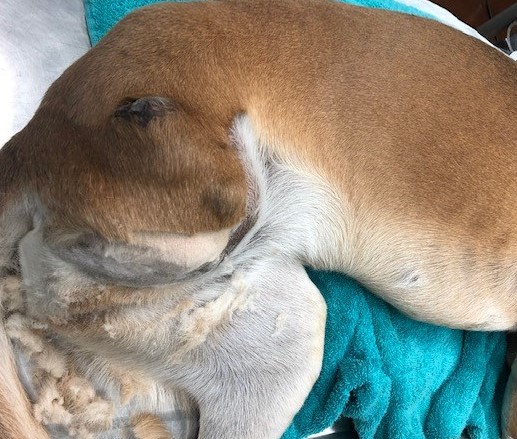
Figure 1: Right femoral amputation site. Multiple nodules are visible on the lateral aspect of the thigh.
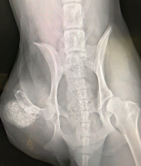
Figure 2: VD radiograph with increased radiodense material on the lateral aspect of the proximal right femur.
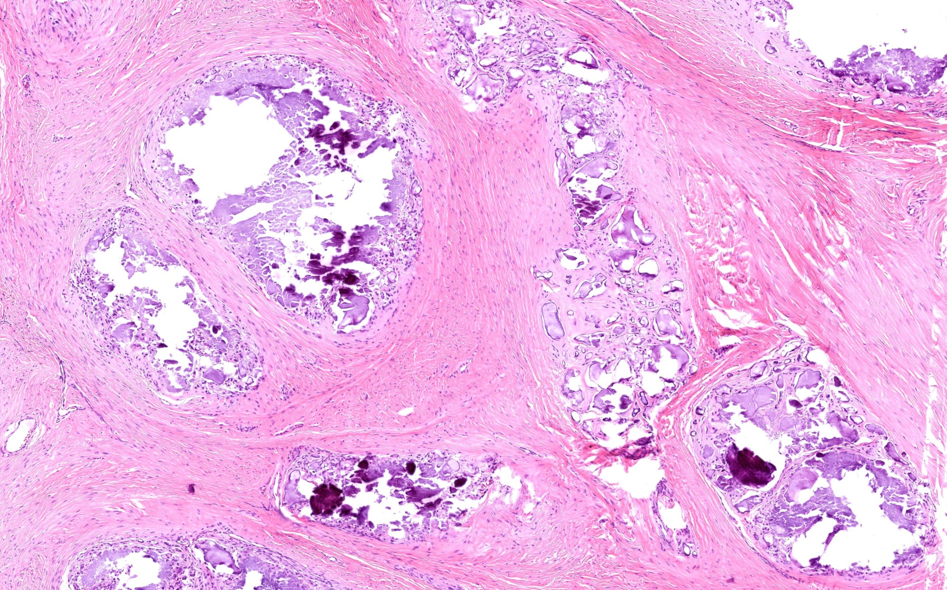
Figure 3: H&E sectin demonstrating multiple granulomas composed of lakes of basophilic granular material (mineral) surrounded and separated by moderate, compressed, lamellar rows of fibroblasts (fibrosis).