Unusual presentation of histoplasmosis in a canine patient
Melanie Landis, DVM, and Julie Piccione,, DVM, MS, DACVP
The clinical pathology section at the Texas A&M Veterinary Medical Diagnostic Laboratory (TVMDL) received a digital cytology submission, with follow-up glass slides submission, of a prostate aspirate from a 7-year-old, male, small-breed canine. According to the referring veterinarian, the patient had a history of weight loss, diarrhea, and tenesmus. Rectal exam revealed an enlarged prostate. Leukocytosis and hypoalbuminemia were found on in-clinic lab work. A fine needle aspirate of the prostate was performed and photographs were taken of the cytologic specimen. TVMDL’s review of the uploaded photographs revealed several cell populations – inflammatory cells (including macrophages, small lymphocytes, and poorly preserved neutrophils) and prostatic epithelial cells. Additionally, the images showed extracellular yeast organisms, as well as yeast phagocytized by macrophages (Figure 1). The yeast were consistent with Histoplasma capsulatum.
Although there have been rare reports of blastomycosis1 in the canine prostate, there are no such reports for histoplasmosis. However, since macrophages migrate to areas of inflammation, and this patient had demonstrable prostatitis, it is reasonable to expect that macrophages with the phagocytized Histoplasma organisms could be detected within the prostate.
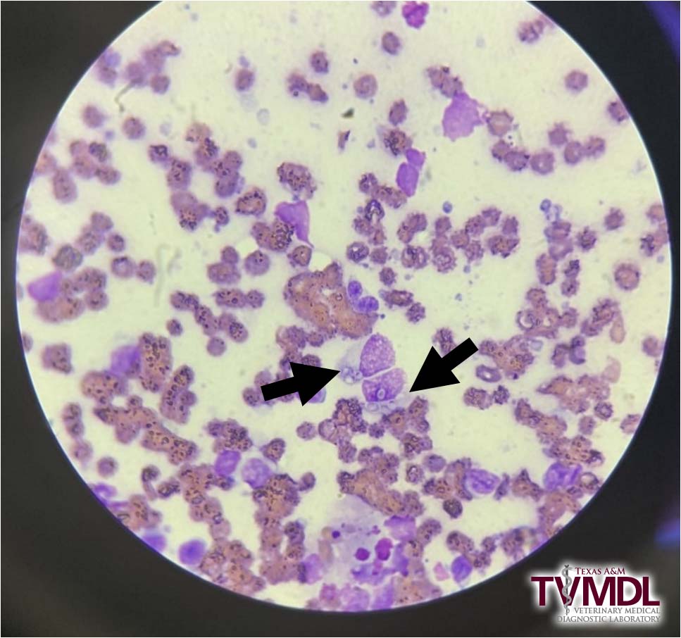
Figure 1: Histoplasma capsulatum (arrows) within macrophages. Image courtesy of referring veterinarian.
While the identification of the fungal organisms, either through cytology, histology or culture, provides the definitive diagnosis, serology can be performed to help determine antibody titers which may be useful in monitoring treatment. Serum from this patient was submitted four days later and antibodies to Histoplasma spp. were detected at a dilution of 1:32 (titer = 32).
TVMDL performs two different serological methods for the detection of fungal antibodies, only one of which provides a titer. The agar gel immune-diffusion (AGID) method is a type of precipitation test where antigen and antibodies diffuse across a gel media. This test is reported as either positive or negative and is a good screening test as it is run with undiluted serum. The complement fixation (CF) test is a more complex test but can yield a titer result. Regardless of method, it should be noted that antibody testing for fungal organisms is not always reliable, especially for Histoplasma.2 In those cases where history, clinical signs, and other diagnostic findings are strongly suggestive of a fungal infection but an antibody test yields negative results, other testing methods (e.g. cytology, histology, culture, antigen testing) should be considered.
As a reminder to our clients, TVMDL’s digital cytology service offers rapid evaluations of microscopic images taken with your smart phone or microscope camera and submitted through the TVMDL Mobile app or on-line at digitalcytology@tvmdl.tamu.edu. The turnaround time is within one hour during normal business hours. This service also includes follow-up submission and evaluation of glass slides, which is included in the price for the digital cytology test.
To learn more about the digital cytology service, visit tvmdl.tamu.edu or call 1.888.646.5623.
References
- KA Arceneaux et al, Blastomycosis in dogs: 115 cases (1980-1995), JAVMA 213:658-664, 1998.
- CE Greene, Infectious Diseases of the Dog and Cat (4th), Chapter 58 – Histoplasmosis, Elsevier, 2012.