Fowl Pox in Houdan chickens
Gabriel Senties-Cue, MVZ, EPAA, MS
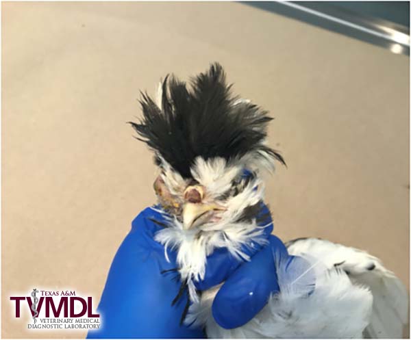
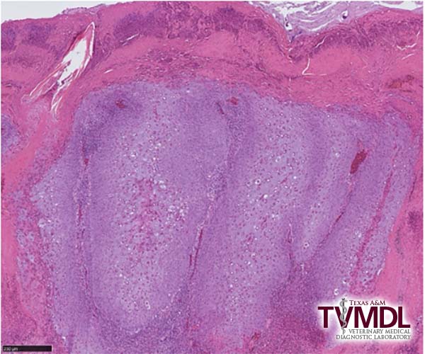
Fowl Pox was diagnosed in three Houdan chickens submitted for diagnostic workup to the Texas A&M Veterinary Medical Diagnostic Laboratory (TVMDL) in Center, Texas. Upon examination, the birds had either one or both eyes severely swollen, due to a large accumulation of caseous exudate, and moderate numbers of dark brown, 3 mm diameter, raised nodules on the eyelids, cere, and skin adjacent to the beak (Figure 1). The palate had a yellowish diphtheritic plaque. The histopathological examination of eyelids and palate revealed characteristic fowl pox diagnostic lesions, including ballooning degeneration and hyperplasia of epithelial cells associated with eosinophilic intracytoplasmic inclusions (Borrel bodies, Figure 2). Additionally, Staphylococcus aureus was isolated from the conjunctiva of the affected eyes, which may have been a secondary infection to fowl pox.
Insects, mainly mosquitoes, serve as mechanical vectors of the fowl pox virus. Fowl Pox or Pigeon Pox live vaccines can be used to immunize birds in areas where the disease is endemic.
For more information about this case, contact Center Resident Director Dr. Gabriel Senties-Cue. To learn more about TVMDL’s test offerings, visit tvmdl.tamu.edu or call one of the agency’s four laboratories.

