• Skip to primary navigation
  • Skip to main content
  • Skip to primary sidebar
  • Skip to footer
  • Locations:
  • Canyon
  • Center
  • College Station
  • Gonzales
  • TVMDL Career Center
  • Contact Us

Texas A&M Veterinary Medical Diagnostic Laboratory

Apple App

download on the Apple App Store
download the app on Google Play

Search

Translate:

  • Find a Form
  • Deliver a Sample
    • Specimen Collection Information
    • Packaging Samples for Shipment
    • Drop Off a Sample
    • Cremation
    • Order Supplies
  • Client Portal
    • Get Test Results
    • SVA Portal
    • Pay a Bill
  • Become a Client
  • Resources
    • TVMDL Bovine Testing Guidance and Reference Material
    • Diagnostic Plans
      • Bovine Syndromic Diagnostic Plans
      • Equine Syndromic Diagnostic Plans
      • Small Ruminant Syndromic Diagnostic Plans
      • Small Animal Syndromic Diagnostic Plans
    • Education Library
    • Case Study Library
    • Electronic Reporting: QR Coding Process
    • CoreOne Resources
  • About Us
    • Locations and Hours
    • Agency Leadership
    • Speaker’s Bureau
    • Texas Pullorum-Typhoid Program
    • TVMDL Mobile app
    • Contact Us
  • Locations
    • College Station Laboratory
    • Canyon Laboratory
    • Center Laboratory
    • Gonzales Laboratory
  • Contact Us

Ochroconosis (Dactylariosis) in birds

August 17, 2020 by Mallory Pfeifer

Ochroconosis (Dactylariosis) in birds
Martin Ficken, DVM

Ochroconosis is a sporadic fungal encephalitis that has been reported in young chickens, turkey poults, quail chicks, and wild birds. It is caused by a dematiaceous, thermophilic fungus, Ochroconis gallopava (Dactylaris gallopava). O. gallopava is a ubiquitous fungus found in hot spring environments, soil, wood, and decaying vegetation. In birds, ochroconosis has been associated with contaminated litter and egg incubators and is presumed to be contracted from inhaling fungal spores from the contaminated environment.1

Clinical signs involve the central nervous system and include tremors, incoordination, torticollis, and paralysis. Death often occurs after a short clinical course. Gross lesions are described as focally extensive circumscribed firm areas of discoloration (gray, red, cream-colored) in the cerebrum and/or cerebellum (Figure 1). In some cases, pulmonary granulomas may also be present. Microscopically, lesions consist of multifocal areas of necrosis infiltrated by heterophils, macrophages, and multinucleated giant cells. Pigmented hyphae are apparent on H&E-stained sections as yellow to light brown, septate, irregularly-branched and 1.2 – 2.4 microns in diameter (Figure 2).

A presumptive diagnosis can be made on histopathological lesions and characteristic pigmented hyphae within the inflammatory lesions. Definitive diagnosis is done by isolation and identification of the organism.2 When the disease occurs, removal of the contaminated litter and/or decontamination of incubators and hatchers by fumigation is recommended.

For more information about poultry testing, contact one of TVMDL’s poultry laboratories in Gonzales or Center. To learn more about TVMDL’s testing options, visit tvmdl.tamu.edu or call 1.888.646.5623.

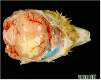
Figure 1 – Turkey poult with cerebral ochroconosis. Notice cream-colored discoloration in right cerebral hemisphere. Photograph courtesy of Dr. H. John Barnes, College of Veterinary Medicine, North Carolina State University.

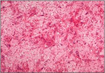
Figure 2 – Turkey poult with cerebral ochroconosis. Notice dark-colored fungal fragments. Photograph courtesy of Dr. H. John Barnes, College of Veterinary Medicine, North Carolina State University.

References

  1. Dykstra MJ, Charlton BR, Chin RP, and Barnes HJ. Fungal Infections in Diseases of Poultry, 13th edition, Wiley-Blackwell, ed. Swayne, DE et al. pp. 1091-1092, 2013.
  2. Gold SE, Glenn AE, and Wyatt RD. Dactylaria in A Laboratory Manual for the Isolation, Identification, and Characterization of Avian Pathogens, 6th Edition, page 75, 2016.

Filed Under: Case Study Tagged With: Avian, bacteria, bacteriology, chicken, fungal, fungus, Gonzales, poultry, TVMDL

Primary Sidebar

Latest Case Studies

  • Historical overview of anthrax in Texas’ livestock population (1974-2022)

    August 11, 2023

  • Juvenile lymphoma in an Angus calf

    July 11, 2023

  • Neurologic Disease due to Bovine Herpesvirus-5 (BHV-5) Infection in a 3-week-old Charolais mix calf

    February 27, 2023

  • Rare case of ocular onchocerciasis in a dog from south Texas

    January 25, 2023

  • Cerebrospinal Nematodiasis (Visceral Larval Migrans) in Birds

    September 13, 2022

Footer

For Employees

  • Employee Email
  • TVMDL Rules & Procedures
  • TVMDL Career Center
  • Emergency Alert Systems
  • AgriLife People Directory
  • AgriLife Administrative Services
  • TAMUS Single Sign On
  • Facebook

State of Texas

  • Texas.gov Portal
  • Texas Veterans Portal
  • Statewide Search
  • Texas Homeland Security
  • Risk, Fraud, & Misconduct Hotline

Policies

  • Privacy and Security Policy
  • Accessibility Policy
  • Texas A&M AgriLife
  • Texas A&M Veterinary Medical Diagnostics Laboratory
  • Texas A&M Forest Service
  • Texas A&M AgriLife Extension Service
  • Texas A&M AgriLife Research

483 Agronomy Rd
College Station, TX 77843