• Skip to primary navigation
  • Skip to main content
  • Skip to primary sidebar
  • Skip to footer
  • Locations:
  • Canyon
  • Center
  • College Station
  • Gonzales
  • TVMDL Career Center
  • Contact Us

Texas A&M Veterinary Medical Diagnostic Laboratory

Apple App

download on the Apple App Store
download the app on Google Play

Search

Translate:

  • Find a Form
  • Deliver a Sample
    • Specimen Collection Information
    • Packaging Samples for Shipment
    • Drop Off a Sample
    • Cremation
    • Order Supplies
  • Client Portal
    • Get Test Results
    • SVA Portal
    • Pay a Bill
  • Become a Client
  • Resources
    • TVMDL Bovine Testing Guidance and Reference Material
    • Diagnostic Plans
      • Bovine Syndromic Diagnostic Plans
      • Equine Syndromic Diagnostic Plans
      • Small Ruminant Syndromic Diagnostic Plans
      • Small Animal Syndromic Diagnostic Plans
    • Education Library
    • Case Study Library
    • Electronic Reporting: QR Coding Process
    • CoreOne Resources
  • About Us
    • Locations and Hours
    • Agency Leadership
    • Speaker’s Bureau
    • Texas Pullorum-Typhoid Program
    • TVMDL Mobile app
    • Contact Us
  • Locations
    • College Station Laboratory
    • Canyon Laboratory
    • Center Laboratory
    • Gonzales Laboratory
  • Contact Us

Fatal Haemonchosis in a Llama

August 17, 2020 by Mallory Pfeifer

Fatal Haemonchosis in a Llama
Erin Edwards, DVM, MS, DACVP and Mindy Borst, LVT

Haemonchosis refers to parasitic infection with Haemonchus contortus, a blood-sucking parasite that lives in the abomasum. This parasite is also known as the barber pole worm due to the macroscopic appearance of the worms. Heavy loads of H. contortus can cause anemia, hypoproteinemia, weight loss, and death. Infection is best described in small ruminants (sheep and goats). Camelids (llamas and alpacas) are also known to be highly susceptible to haemonchosis.

Recently, a deceased, 2-year-old llama from the Brazos Valley was submitted to the Texas A&M Veterinary Medical Diagnostic Laboratory (TVMDL) for necropsy. This llama had a history of parasitic resistance and severe anemia that required blood transfusion. Postmortem examination revealed a thin body condition, severe pallor of the ocular and oral mucus membranes, subcutaneous edema, tricavitary effusions, and pallor of the internal organs. The third gastric compartment (C3), which is equivalent to the abomasum in ruminants, contained tremendous amounts of parasites that were identified as H. contortus (Figure 1). The parasite burden in this llama was responsible for the other noted necropsy findings and was determined to be the primary cause of death.

H. contortus thrives in warm, humid environments such as Texas. Haemonchosis is a major problem in small ruminants and camelids in tropical environments, including other parts of the southeastern United States. This parasite has become increasingly more resistant to anthelmintics and infections are often hard to clear, as demonstrated in this case. For more information on developing strategies to reduce resistance and manage this disease, visit the Texas A&M AgriLife Extension website.

For more information about this case, contact Dr. Erin Edwards, assistant anatomic pathology section head, or Mindy Borst, clinical pathology laboratory supervisor. To learn more about TVMDL’s test offerings, visit tvmdl.tamu.edu or call 1.888.646.5623.

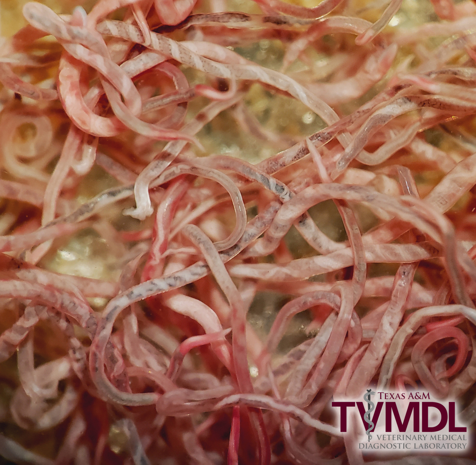
Figure 1: Haemonchus contortus nematodes that were recovered from the third gastric compartment of a 2-year-old llama. Note the twisted appearance of some, which is why this parasite is also known as the barber pole worm.

Reference:

Edwards EE, Garner BC, Williamson LH, Storey BE, and Sakamoto K. Pathology of Haemonchus contortus in New World camelids in the southeastern United States: a retrospective review. J Vet Diagn Invest. 2016.

Filed Under: Case Study Tagged With: camelid, clinical pathology, college station, exotic, histopathology, llama, parasitology, pathology, TVMDL

Primary Sidebar

Latest Case Studies

  • Neurologic Disease due to Bovine Herpesvirus-5 (BHV-5) Infection in a 3-week-old Charolais mix calf

    February 27, 2023

  • Rare case of ocular onchocerciasis in a dog from south Texas

    January 25, 2023

  • Cerebrospinal Nematodiasis (Visceral Larval Migrans) in Birds

    September 13, 2022

  • Spironucleosis (Hexamitiasis) in Quail

    August 9, 2022

  • Endocarditis in a White-Tailed Deer Caused by Trueperella pyogenes

    August 9, 2022

Footer

For Employees

  • Employee Email
  • TVMDL Rules & Procedures
  • TVMDL Career Center
  • Emergency Alert Systems
  • AgriLife People Directory
  • AgriLife Administrative Services
  • TAMUS Single Sign On
  • Facebook

State of Texas

  • Texas.gov Portal
  • Texas Veterans Portal
  • Statewide Search
  • Texas Homeland Security
  • Risk, Fraud, & Misconduct Hotline

Policies

  • Privacy and Security Policy
  • Accessibility Policy
  • Texas A&M AgriLife
  • Texas A&M Veterinary Medical Diagnostics Laboratory
  • Texas A&M Forest Service
  • Texas A&M AgriLife Extension Service
  • Texas A&M AgriLife Research

483 Agronomy Rd
College Station, TX 77843