Leishmania mexicana diagnosed in feline ear biopsy specimens – A classic entity in Texas cats
Erin Edwards, DVM, MS, DACVP
Two biopsy specimens from the pinnae of a 6-year-old, domestic shorthair cat from east-central Texas were submitted to the Texas A&M Veterinary Medical Diagnostic Laboratory (TVMDL) for histologic examination. This cat was reported to have red, raised, hemorrhagic masses on both pinnae which were removed with surgical excision. Histologically, there were three separate masses identified within the received biopsy specimens. These areas consisted of large, ulcerated nodules of severe granulomatous inflammation. Numerous small, protozoal amastigotes were seen within the cytoplasm of macrophages. These organisms were histologically consistent with Leishmania spp. and, based on the signalment and lesion location, were diagnosed as Leishmania mexicana.
The lesions in this case exemplify cutaneous leishmaniasis that is seen in Texas cats caused by L. mexicana. There are many species of Leishmania across the world which can cause cutaneous, mucocutaneous, and visceral infection. L. mexicana is endemic in Texas and causes the cutaneous form of this disease. Cases have been diagnosed across Texas, including as far north as the Dallas-Fort Worth area. Anecdotally, there have been many cases in recent years from parts of the Brazos Valley.
In reported cases, lesions associated with L. mexicana have been restricted to the skin. Infected cats present with one or more cutaneous nodules that are classically located on the pinnae. Other areas of skin are less commonly affected, typically on the face. The aural and facial distribution of these lesions reflects the site predilection for vector insect bites. Leishmania spp. are transmitted by sandflies. Humans and various animal species can be infected, though cats seem to be more susceptible to L. mexicana. Outdoor cats are at increased risk of infection. Immunocompetent cats are susceptible and an association with FeLV/FIV infection has not been demonstrated. The cat in this case was negative for both FeLV and FIV.
There are other important differential diagnoses for masses on the pinnae of cats which can clinically resemble cutaneous leishmaniasis, including mast cell tumor and other types of neoplasia. Diagnosis of leishmaniasis can be achieved with cytology and/or biopsy. The protozoal organisms characteristically contain a bar-shaped kinetoplast that is oriented perpendicular to the protozoal nucleus. This feature is best visualized with cytology. In rare cases, cutaneous lesions have been reported to spontaneously resolve, though this process may be prolonged. Surgical excision is often locally curative, though some reports have described mass recurrence.
For more information on this case, contact Dr. Erin Edwards, pathologist at the College Station laboratory. To learn more about TVMDL’s test offerings, visit tvmdl.tamu.edu.
References:
- Trainor KE, Porter BF, et al. Eight Cases of Feline Cutaneous Leishmaniasis in Texas. Vet Pathol. 47(6): 1076-1081.
- Minard HM, Daniel AK, et al. Pathology in Practice. J Am Vet Med Assoc. 2017. 251(1): 57-59.
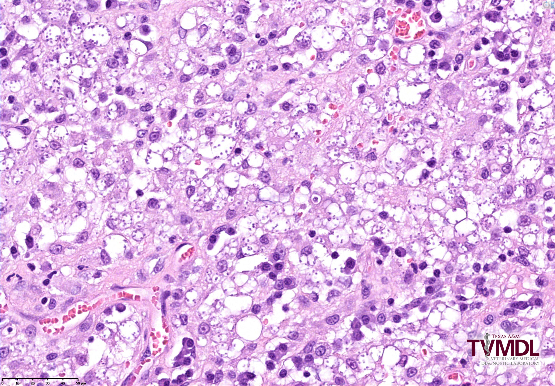
Figure 1: Histopathology of a mass on the pinna of a cat from Texas. This image shows sheets of macrophages that contain numerous small, protozoal amastigotes consistent with Leishmania mexicana.