Testing Options for Anaplasma marginale in Cattle
Melanie T. Landis, DVM, MBA
Anaplasma marginale is a rickettsial bacterium that invades the red blood cells (RBCs) of ruminants, primarily cattle, and is considered the most common tick-borne infection of cattle. In addition to tick vectors (Dermacenter spp., Rhipicephalus spp.), biting flies and blood-tainted fomites can also transmit this organism. Anaplasma marginale is typically a clinical disease of adult cattle. Calves infected with the organisms usually show no, or only mild, clinical signs of disease. However, once recovered, these calves are considered persistently infected, and can serve as a reservoir host. In adult cattle, clinical signs in the acute phase of infection include sudden death, abortion, pale and/or icteric mucus membranes, fever, dehydration, depression, and anorexia and are in part related to the number of RBCs infected. During the acute phase parasitemia is pronounced, with the organism infecting between 20-70% of the RBCs, but quickly drops following onset of clinical signs. The infection results in extra-vascular hemolysis, so hemoglobinemia and hemoglobinuria are not seen, although regenerative anemia eventually develops. Additionally, bilirubinemia and bilirubinuria may be elevated during the convalescent period and weight loss can to be noted. If adult cattle recover, they too, are considered persistently infected reservoirs. Further, parasitemia may recur if the animal is immunosuppressed or develops a co-infection.1
There are several options for testing for Anaplasma, and selection is partly dependent on the stage of infection, or whether the test is used for diagnosing or screening cattle:
- Hemoparasite exam of blood smears is best utilized during the acute phase of infection. Once clinical signs are evident, blood smear examination should occur within a matter of days to optimize the likelihood of finding the Anaplasma organism within RBCs. Examination of blood slides during the convalescent phase typically yields negative results. Blood should be collected in an EDTA tube and thin blood smears (i.e. air dried slides) prepared at the time of collection. Wright-Giemsa stain, as well as Diff-Quick®, can be utilized for staining.
- qPCR (real time polymerase chain reaction) testing utilizes EDTA whole blood or fresh tissues to look for the DNA of the Anaplasma qPCR is best at detecting acute infections when bacteremia is still present, but may still be able to detect some infections after visual identification of the organism is no longer present on a blood smear.
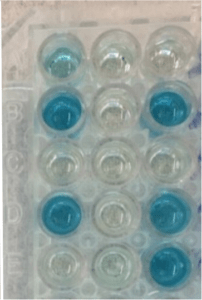
- cELISA (competitive ELISA) testing is an antibody test that utilizes serum and is a good method to help detect carriers, but may yield negative results in early acute infections. Serum is best submitted off the clot, and hemolyzed samples cannot be tested. This test provides a relatively easy method for screening an entire herd. Results are reported as positive or negative utilizing the calculation of % inhibition, which is based on the optical density of the sample as detected by an automated cELISA plate reader. Low % inhibition correlates with negative results and is visually demonstrated by dark color change of the sample.
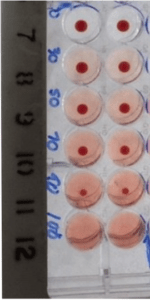
- Complement Fixation (CF) testing is an antibody test that runs on the premise that if antibodies are present in the sample, they will fix complement and prevent hemolysis of added antibody-sensitized sheep blood. When requested, this test is referred to the National Veterinary Services Laboratory (NVSL) in Ames, IA. Serum is best submitted off the clot, and hemolyzed samples cannot be tested. The CF test has shown a high degree of variability in sensitivity. Further, this testing method is not as good at detecting carrier states as cELISA testing. Finally, although once thought to be most useful in detecting acute infections, it is now unclear if CF testing can detect acute infections any sooner than other antibody tests. Therefore, while CF tests will yield a titer, unless it is required for export purposes, this testing method is no longer recommended for routine diagnostics.2

Anaplasma marginale on blood smear – Giemsa stain. Photograph curtesy of Julie Piccione, DVM, MS, DACVP

cELISA – Well A-1 is positive control; Well B-1 is negative control. Wells with color intensity greater than well A-1 are negative

Complement fixation – The top two pairs of wells are considered positive as complement has fixed the antibodies present in the sample and prevented hemolysis of the sheep blood.
To learn more about TVMDL’s bovine testing options, visit tvmdl.tamu.edu. TVMDL employs veterinary diagnosticians who are available for consultation on test recommendations and result interpretation. Call TVMDL’s College Station or Amarillo laboratories to schedule a diagnostic consultation.
REFERENCES:
- Palmer, G. Infectious Causes of Hemolytic Anemia – Anaplasmosis, Large Animal Internal Medicine, 4th, BP Smith, DVM, DACVIM, editor. Mosby Elsevier ©2009, pp 1155-1157
- OIE Terrestrial Manual 2018, Chapter 3.4.1 – Bovine anaplasmosis. oie.int/fileadmin/Home/eng/Health_standards/tahm/3.04.01_BOVINE_ANAPLASMOSIS.pdf