Ocular coccidiomycosis in a puppy
Erin Edwards, DVM, MS, DACVP
Last summer, ocular coccidiomycosis was diagnosed via histopathology in a 12-week-old Great Pyrenees puppy. The puppy was from Brewster county in West Texas. This puppy was clinically diagnosed with juvenile glaucoma in the right eye and a previous history of possible unknown trauma was speculated. The affected eye was enucleated and sent to the Texas A&M Veterinary Medical Diagnostic Laboratory (TVMDL) for histopathologic examination (Figure 1).
Histopathology revealed that the glaucoma in this case was secondary to a severe, pyogranulomatous panophthalmitis. Inflammation extended into all parts of the eye and was most severe along the posterior portions of the globe. Scattered throughout the inflammatory exudate and often within the center of small pyogranulomas were frequent fungal spherules consistent with Coccidioides spp. These spherules rarely exhibited central endosporulation (Figure 2). In addition to glaucoma, other secondary changes included retinal detachment and corneal neovascularization.
Coccidiomycosis is a fungal infection that can be caused by Coccidiodes posadasii or C. immitis. The latter is typically geographically restricted to the San Joaquin Valley in California where the disease is often known as Valley Fever. C. posadasii is endemic across the southwestern United States, including Texas, and parts of Mexico. Cases are frequently diagnosed at TVMDL. Dogs are the most frequently affected species and camelids are also highly susceptible to this disease. Of note, humans are also susceptible to infection.
Infection with coccidiomycosis occurs following inhalation of arthrospores from contaminated soil. This disease is seen most often in young, outdoor sporting or working dogs, likely associated with digging behaviors that stir up and aerosolize arthrospores. The infected puppy in this case was surprisingly young. Following inhalation, infection is first established in the lungs and infected animals often show signs of respiratory disease. In a few cases, the infection becomes disseminated to other sites. The bones, skin, and eyes are the most commonly affected distant sites, though many viscera can also be involved. In some cases, such as the currently discussed case, infected patients may present for clinical signs associated with infection at distant sites without showing signs of pneumonia.
The TVMDL case study library contains a few other cases of coccidiomycosis that should be explored for more information on this disease. These cases demonstrate the variety of case presentations seen at TVMDL.
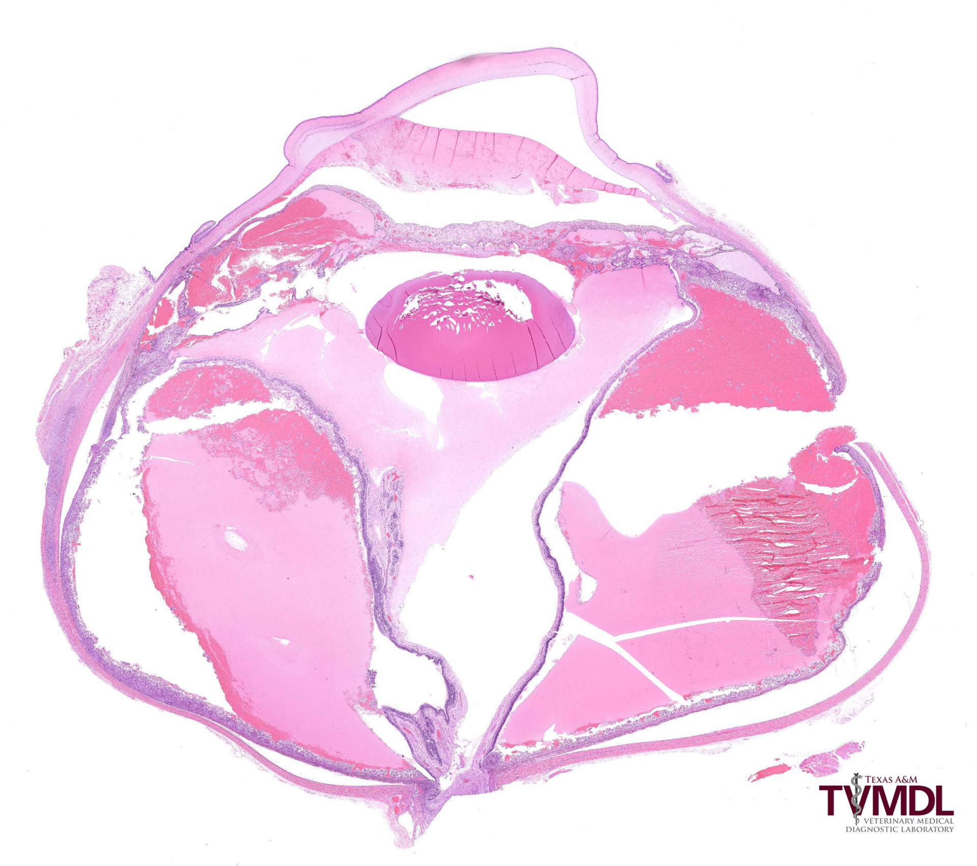
Figure 1: A low-power image of the enucleated globe showing buphthalmia, retinal detachment, and severe intraocular hemorrhage and inflammation.
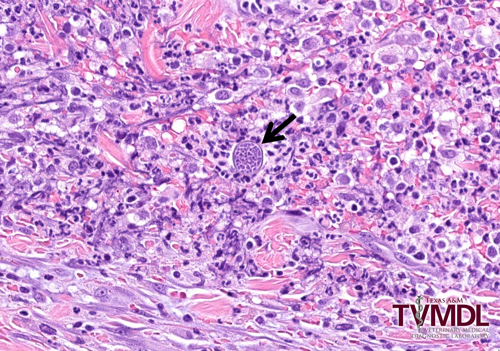
Figure 2: This image is from an area of pyogranulomatous scleral inflammation. In the center there is a Coccidioides spp. fungal spherule with endosporulation.