Horse Diagnosed with Equine Botryomycosis
Kayla Tomaso and Erin Edwards, DVM, MS, DACVP
Tissue samples from two ear masses on a 3-year-old American Quarter Horse were submitted to the Texas A&M Veterinary Diagnostic Lab (TVMDL) in College Station for histopathology and culture. Masses from both ears were histologically similar, consisting of severe, chronic, pyogranulomatous dermatitis. There was formation of discrete pyogranulomas with central aggregates of Splendore-Hoeppli material and intralesional bacteria (Figure 1). The culture yielded mixed bacterial growth with a predominance of Staphylococcus aureus (Figure 2). These results were consistent with the diagnosis of botryomycosis.
Botryomycosis, also called bacterial pseudomycosis due to its clinical and histological resemblance to fungal infections, is an uncommon granulomatous skin condition caused by non-filamentous bacteria. Botryomycosis is most often caused by Staphylococcus aureus; however, Pseudomonas aeruginosa, Streptococcus spp., and a few other bacteria have rarely been implicated. S. aureus can be a commensal organism or an opportunistic pathogen and the specific factors that elicit the pseudomycetomatous inflammation are unclear. Histologic lesions resemble those of actinomycotic and eumycotic fungal mycetomas and are similar to lesions of lumpy jaw, caused by Actinomyces bovis, and wooden tongue, caused by Actinobacillus lignieresii, in cattle.
This disease can be seen in many animal species. Most cases are associated with a cutaneous wound from local trauma, castration sites being a common location in horses. Lesions are typically solitary but in rare cases there can be disseminated masses with widespread cutaneous involvement. Therapy for botryomycosis typically includes surgical excision. For resistant or disseminated cases, treatment may require systemic antimicrobial treatment.
For more information about this case, contact Kayla Tomaso, bacteriology technician, or Dr. Erin Edwards, veterinary pathologist, at the College Station laboratory. To learn more about TVMDL’s testing services, visit tvmdl.tamu.edu or call 1.888.646.5623.
References:
– M Hargis and S Myers. The Integument. In: JF Zachary, ed. Pathologic Basis of Veterinary Disease. 6th ed. St. Louis, MO: Elsevier, Inc; 2017: 1077.
– DW Scott and WH Miller, Jr. Equine Dermatology. Maryland Heights, MO: Elsevier Saunders; 2011: 161-162.
– J Songer and KW Post. Veterinary Microbiology Bacterial and Fungal Agents of Animal Disease. St. Louis, MO: Elsevier Saunders; 2005: 38, 156.
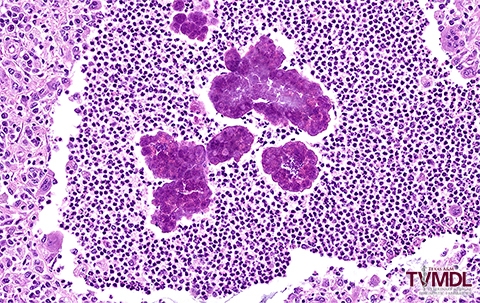
Figure 1: Histopathology image showing a pyogranuloma with large aggregates of Splendore-hoeppli material and small amounts of central bacterial cocci.
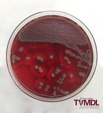
Figure 2: Blood agar plate with Staphylococcus aureus and mixed bacterial growth.