Chromatophoromas in lacertids
By Gaya Balamayooran, DVM, PhD, DACVP and Josué Diaz-Delgado, DVM, MS, PhD, DACVP
A 76 gram, 9-year-old female knight anole (Anolis equestris) lizard from a zoological collection was euthanized due to poor prognosis after recurrence of a skin neoplasm on the lateral body that was incompletely excised 9 months prior. The neoplasm extended to the head at the time of euthanasia. A mixed chromatophoroma was diagnosed histologically at the Texas A&M Veterinary Medical Diagnostic Laboratory (TVMDL) based on the presence of melanophores (melanin) and birefringent iridophores (Figures 1A and 1B). This neoplasm had a mitotic count of 10 per 10 high power fields. Metastasis was not observed in distant organs, including the liver and lung, among the limited tissues submitted for histologic examination.
A 436 gram, 7-year-old, client-owned, female bearded dragon (Pogona vitticeps) presented for an approximately 1 cm in diameter raised nodule on the middle of the back, which was first noticed about 6 months prior. The mass had expanded quickly over the last 2 months. A punch biopsy was submitted to TVMDL for histopathologic examination. Microscopically, the mass had cytohistomorphologic features consistent with a mucinous melanophoroma (Figures 2A and 2B). In this case, tumor cells extended to one of the lateral surgical margins. Since local invasion and metastasis have been reported in melanophoromas in bearded dragons, close monitoring of this patient was advised. That specific margin was widened shortly after. No recurrence has been reported at this time.
Lizards have a median incidence of neoplastic disease compared with snakes, turtles, and tortoises, and the hematopoietic, cutaneous, and hepatic systems are most commonly affected.1 Chromatophoromas, including melanophoromas, xanthophoromas and iridophoromas, are tumors of pigment-producing cells of the skin and are relatively common in reptiles.2,3 Iridophoroma has been described in bearded dragon, savannah monitor, and veiled chameleon. To the best of our knowledge, a mixed tumor with melanin and birefringent pigments has not been reported in knight anole lizards. Mixed chromatophoroma has been reported in snakes.4 Within lacertid melanophoromas, a “mucinous” variant, based on cytohistomorphologic and tinctorial properties, has only been recognized in few bearded dragons.2,3 This subtype was associated with poorer prognosis due to local invasiveness.2,3
For more information about this case, contact Dr. Díaz-Delgado or Dr. Balamayooran, pathologists at the College Station laboratory. To learn more about TVMDL’s test offerings, visit tvmdl.tamu.edu or call the TVMDL laboratory nearest to you.

Figure 1. (A) Subgross microphotograph of mixed chromatophoroma in a knight anole lizard (Anolis equestris). (B) The mixed chromatophoroma is composed of neoplastic spindle cells with varying amounts of birefingent pigment (blue arrow) and melanin pigment (dark brown arrow). (C) Mitotic figures (arrows) in neoplastic cells. (D) Neoplastic cells with birefingent pigment under polarized light. Hematoxylin-Eosin stain.
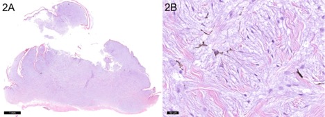
Figure 2. (A) Subgross microphotograph of mucinous melanophoroma in a bearded dragon (Pogona vitticeps) (B). The mucinous melanophoroma is composed of melanized and amelanotic stellate to spindle cells accompanied by extracellular mucinous fluid and collagen fibers.
References
- Christman, J., Devau, M., Wilson-Robles, H., Hoppes, S., Rech, R., Russell, K. E., & Heatley, J. J. (2017). Oncology of reptiles: diseases, diagnosis, and treatment. Veterinary Clinics: Exotic Animal Practice, 20(1), 87-110.
- Heckers, K. O., Aupperle, H., Schmidt, V., & Pees, M. (2012). Melanophoromas and iridophoromas in reptiles. Journal of comparative pathology, 146(2-3), 258-268.
- Heckers, K. O., Aupperle, H. (2014). Pigment-forming tumors in reptiles: light regime and its dark sides. In: Proceedings of the Association of Reptilian and Amphibian Veterinarians, 21th Annual Conference, Orlando, Florida, USA, Association of Reptilian and Amphibian Veterinarians. 18-24 October, 2014, 31-35.
- Muñoz-Gutiérrez, J. F., Garner, M. M., & Kiupel, M. (2016). Cutaneous chromatophoromas in captive snakes. Veterinary Pathology, 53(6), 1213–1219