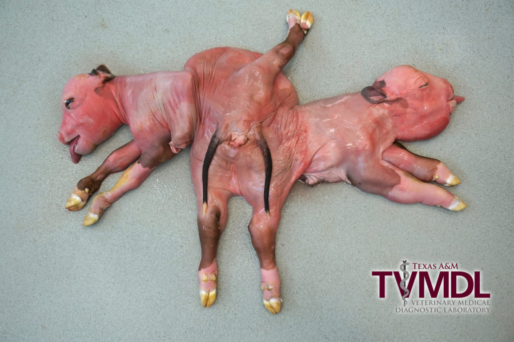
Necropsy of rare bovine conjoined twins
By Erin Edwards, DVM, MS, DACVP and Gaya Balamayooran, DVM, PhD, DACVP
Recently, bovine conjoined twin calves were submitted to the Texas A&M Veterinary Medical Diagnostic Laboratory (TVMDL). Both the dam and sire were angus crosses from a herd of 250 head in the eastern region of Texas. There had been no prior occurrences of conjoined twins or other congenital abnormalities on the property. The dam had previously delivered multiple normal calves. She was noted to be in dystocia and a ranch hand helped pull the conjoined twin fetus. The specimen was taken to TVMDL where necropsy was performed.
The twins were both female and were joined at the pelvis with the heads facing in opposite directions. The animal had two heads, four forelimbs, three hindlimbs, two tails, and one umbilicus. One hindlimb consisted of two fused limbs and was positioned dorsally above the tails. The two hindlimbs located ventrally were attached to two discernible acetabular fossa, but a discernible acetabulum was not present attached to the dorsal fused hindlimb. No anus was identified. There were two adjacent vulvar openings which were separate externally but joined into a common vagina. In one twin, two uterine horns opened into two cervices and entered the vagina separately, whereas in the other twin, both uterine horns blindly ended at the vaginal wall without a cervix. There was one spiral colon and one distal colon which emptied into the vagina. All other organs were present in duplicate. The kidneys from each twin entered into separate urinary bladders (each bladder was supplied by a ureter from each fetus). The liver of one twin was over twice the size of that from the other. One twin had severe hydrocephalus.
Conjoined twins can be classified depending on the location of their anatomical fusion, and there are many types. In this case, the conjoined twins are classified as ischiopagus tripus based on the pelvic union with 3 hind limbs.
Conjoined twins are rare in animals. Among domestic species, cattle are the most commonly affected. There are no recent reports on the incidence of this anomaly in cattle. Older reports, however, report an incidence of 1 in every 100,000 births. The vast majority of these are considered spontaneous and a cause is seldom identified. Conjoined twins are believed to be the result of partial splitting of a fertilized egg. There are various causes of other congenital anomalies and malformations in cattle; however, reports of direct associations with conjoined twins are unsubstantiated. Possible causes could include genetic defects, consumption of teratogenic toxins by the dam, and viral infection. In the United States, viruses that have been shown to induce fetal anomalies in ruminants include bovine viral diarrhea virus (BVD), Cache Valley fever virus, and bluetongue virus. Testing for these viruses is recommended in any diagnostic workup of conjoined twins. A viral cause was not detected in this case.
For more information about this case, contact Dr. Erin Edwards or Dr. Gaya Balamayooran, veterinary pathologists at the College Station laboratory. To learn more about bovine testing, visit tvmdl.tamu.edu or call 1.888.646.5623.
References:
- Agerholm, J.S., Hewicker-Trautwein, M., Peperkamp, K., and Windsor, P.A. (2015). Virus-Induced Congenital Malformations in Cattle. Acta Vet Scand, 57(54), 1-14.
- Hiraga, T., and Dennis, S.M. (1993). Congenital Duplication. Vet Clin N Am-Food A, 9(1), 145-161.
- Kumar, S., Pandey, A.K., Kushwaha, R.B., Sharma, U., and Dwivedi, D.K. (2014). Dystocia Due to Conjoined Twin Monster in a Cow. Indian J Anim Reprod, 35(1), 54-56.
- Vanderzon, D.M., Partlow, G.D., Fisher, K.R.S., and Halina, W.G. (1998). Parapagus Conjoined Twin Holstein Calf. Anat Rec, 251, 60-65.
