Contagious Ecthyma Diagnosed in a Young Goat
Erin Edwards, DVM, MS, DACVP, Andrés de la Concha-Bermejillo, DVM, PhD, Pam Ferro, MS, PhD
The body of a 3-month-old goat was submitted to the Texas A&M Veterinary Medical Diagnostic Laboratory (TVMDL) in College Station. This goat had been purchased 3 weeks prior. The goat was euthanized due to sudden onset of neurological signs and was also noted to have a crusty lesion on the lip. Gross necropsy confirmed there to be an exophytic, crusty mass on the commissure of the lip (Figure 1). Other necropsy lesions were minor and histopathology was pursued.
The lip lesion was histologically consistent with contagious ecthyma, also known as orf or soremouth. This mass consisted of an area of proliferative chelitis with subcorneal vesicles and pustules. In a few areas epithelial cells had ballooning degeneration and contained intracytoplasmic, eosinophilic, viral inclusion bodies consistent with parapoxvirus (Figure 2). There was also evidence of secondary bacterial infection. A section of this lip mass was submitted for parapoxvirus rtPCR. High levels of parapoxvirus DNA were detected, confirming the diagnosis of contagious ecthyma. This goat was also diagnosed with polioencephalomalacia as the cause of the neurologic disease.
Contagious ecthyma is caused by a parapoxvirus and results in raised, crusty lesions which are most commonly found at mucocutaneous junctions, particularly around the mouth. In more severe cases, the skin of other areas such as the eyelids, feet, vulva, and udder may also have lesions. This disease is most commonly seen in small ruminants though camelids and rarely other ruminants can also be affected. This virus is zoonotic and humans can be infected after contact with affected lesions. The disease is usually self-limiting and immunocompromised individuals are more at risk. The disease usually runs its course in 3–4 weeks; however, prolonged infections and secondary bacterial infections or myiasis of affected areas may occur. Affected animals should be handled with gloves with care taken to avoid contact with obvious lesions.
For more information about this case, contact Dr. Edwards or Dr. de la Concha-Bermejillo, pathologists, or Dr. Ferro, molecular diagnostics section head. To learn more about TVMDL’s testing options, visit tvmdl.tamu.edu or call 1.888.646.5623.
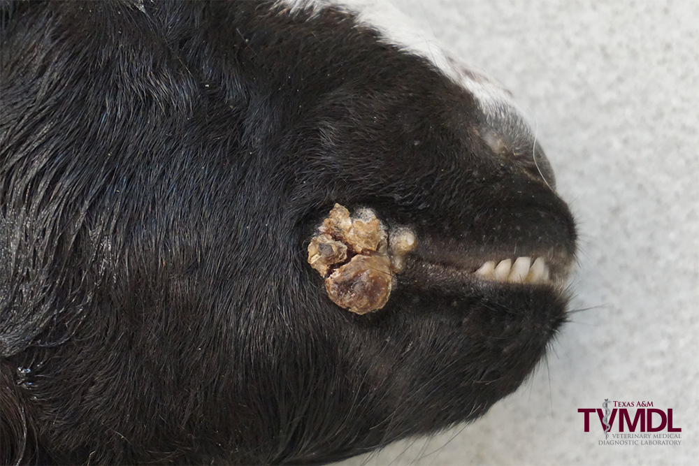
Figure 1: This image shows a crusty, exophytic mass at the lip commissure in a goat with contagious ecthyma.
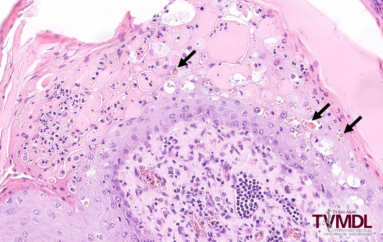
Figure 2: Histopathology of the lip mass in this goat showing epithelial cell degeneration with many intracytoplasmic viral inclusion bodies, some of which are indicated with arrows.
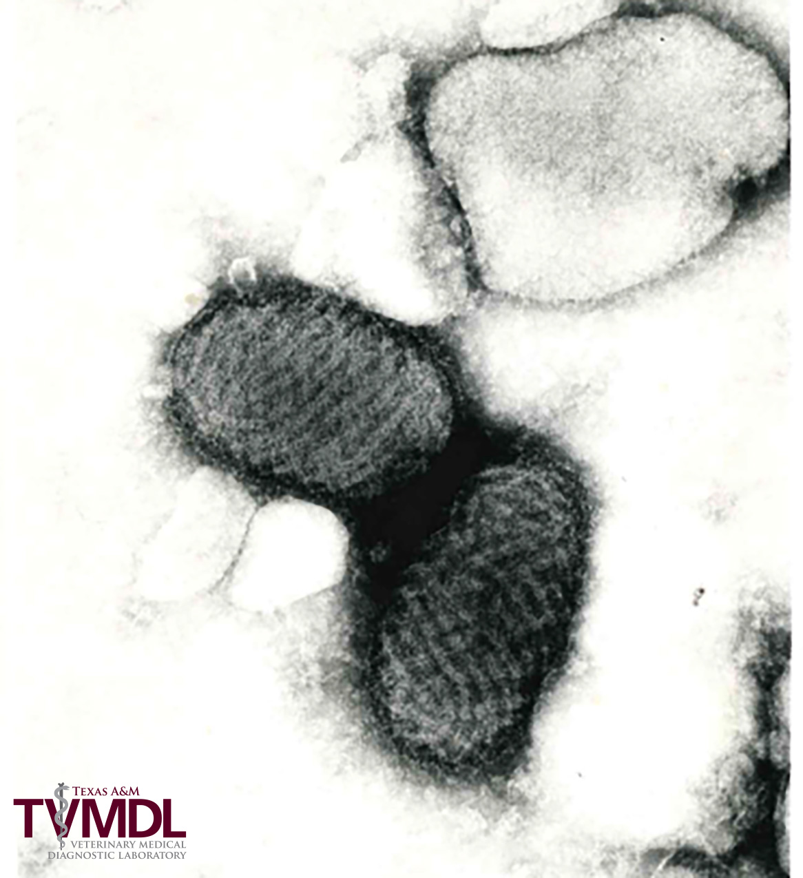
Figure 2: Negative stained parapoxvirus (Orf virus, TEM image)
References:
- de la Concha-Bermejillo A. Orf (Contagious ecthyma). In: Blackwell’s Five-Minute Veterinary Consult. Ed. by CCL Chase. Wiley Blackwell. Second Edition. pp 564-565, 2017
- de la Concha-Bermejillo A, et al. Severe persistent orf in young goats. J Vet Diagn Invest. 2003 Sep;15(5):423-31