• Skip to primary navigation
  • Skip to main content
  • Skip to primary sidebar
  • Skip to footer
  • Locations:
  • Canyon
  • Center
  • College Station
  • Gonzales
  • TVMDL Career Center
  • Contact Us

Texas A&M Veterinary Medical Diagnostic Laboratory

Apple App

download on the Apple App Store
download the app on Google Play

Search

Translate:

  • Find a Form
  • Deliver a Sample
    • Specimen Collection Information
    • Packaging Samples for Shipment
    • Drop Off a Sample
    • Cremation
    • Order Supplies
  • Client Portal
    • Get Test Results
    • SVA Portal
    • Pay a Bill
  • Become a Client
  • Resources
    • TVMDL Bovine Testing Guidance and Reference Material
    • Diagnostic Plans
      • Bovine Syndromic Diagnostic Plans
      • Equine Syndromic Diagnostic Plans
      • Small Ruminant Syndromic Diagnostic Plans
      • Small Animal Syndromic Diagnostic Plans
    • Education Library
    • Case Study Library
    • Electronic Reporting: QR Coding Process
    • CoreOne Resources
  • About Us
    • Locations and Hours
    • Agency Leadership
    • Speaker’s Bureau
    • Texas Pullorum-Typhoid Program
    • TVMDL Mobile app
    • Contact Us
  • Locations
    • College Station Laboratory
    • Canyon Laboratory
    • Center Laboratory
    • Gonzales Laboratory
  • Contact Us

Heterobilharzia americana Infection in a Dog

May 28, 2019 by Mallory Pfeifer

Heterobilharzia americana Infection in a Dog
Jay Hoffman, DVM, PhD

Small intestinal biopsy samples from a two-year-old German Shepherd dog were submitted to the Texas A&M Veterinary Medical Diagnostic Laboratory (TVMDL) in College Station. The clinical history indicated chronic diarrhea for the last six months, normal blood parameters and no parasites noted on fecal floatation. When the biopsies were examined there were numerous white nodules noted in the wall of the small intestine (Figure 1). Microscopically these nodules contained numerous trematode ova, approximately 45-90 um in diameter, located within the deep muscular layers of the small intestine. Surrounding the trematode ova were thick strands of fibrous connective tissue entrapping infiltrates of multinucleated giant cells, macrophages, lymphocytes, and plasma cells. The trematode ova were histologically compatible with Heterobilharzia americana.

Heterobilharzia americana is a trematode parasite endemic to the Gulf Coast region of Texas.  The adults are blood parasites and live in the mesenteric veins where mating occurs. The female produces thousands of ova that lodge in the enteric venules, secrete proteolytic enzymes and eventually gain access to the lumen of the small intestine. Other ova become lodged in the wall of the small intestine and induce granulomatous inflammation. Some of the ova bypass the enteric venules and become lodged in the liver, lymph nodes, and pancreas and induce inflammation. Once the ova are passed in the feces, they hatch and form miricidia once they contact water. These miricidia penetrate the soft tissues of freshwater snails where they reproduce and release free-swimming cercariae. The cercariae can invade the skin or mucous membranes of the definitive host (dogs or wild mammals) and gain access to the bloodstream, migrate to the liver, mature, and migrate to the mesenteric vessels to complete the life cycle.  The clinical signs observed in infected animals include weight loss, hematochezia, melena, diarrhea, vomiting, and lethargy. It is likely that the clinical signs are primarily caused by the inflammation induced by the trematode ova.

For more information about this case, contact Dr. Jay Hoffman, veterinary pathologist, at the College Station laboratory. To learn more about TVMDL’s test offerings, visit tvmdl.tamu.edu or call 1.888.646.5623.

Photomicrograph of numerous trematode ova surrounded by granulomatous inflammation and fibrous connective tissue.

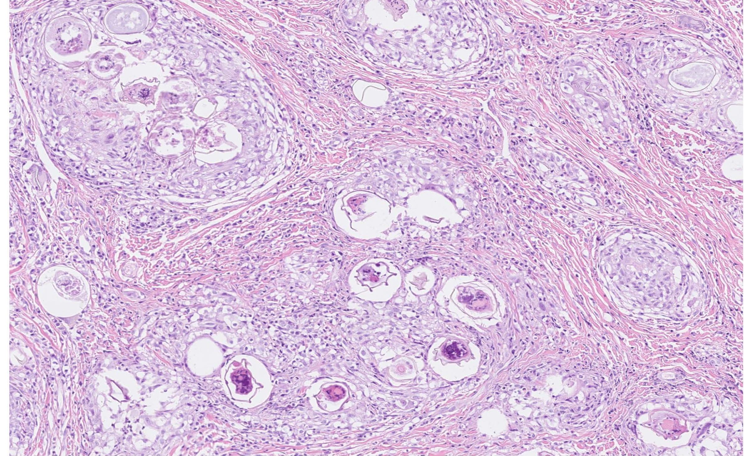
Figure 1. Small intestine (deep muscular layers) from a dog. Numerous trematode ova surrounded by granulomatous inflammation and fibrous connective tissue.

Filed Under: Case Study Tagged With: canine, college station, dog, German Shepherd, histopathology, pathology

Primary Sidebar

Latest Case Studies

  • Neurologic Disease due to Bovine Herpesvirus-5 (BHV-5) Infection in a 3-week-old Charolais mix calf

    February 27, 2023

  • Rare case of ocular onchocerciasis in a dog from south Texas

    January 25, 2023

  • Cerebrospinal Nematodiasis (Visceral Larval Migrans) in Birds

    September 13, 2022

  • Spironucleosis (Hexamitiasis) in Quail

    August 9, 2022

  • Endocarditis in a White-Tailed Deer Caused by Trueperella pyogenes

    August 9, 2022

Footer

For Employees

  • Employee Email
  • TVMDL Rules & Procedures
  • TVMDL Career Center
  • Emergency Alert Systems
  • AgriLife People Directory
  • AgriLife Administrative Services
  • TAMUS Single Sign On
  • Facebook

State of Texas

  • Texas.gov Portal
  • Texas Veterans Portal
  • Statewide Search
  • Texas Homeland Security
  • Risk, Fraud, & Misconduct Hotline

Policies

  • Privacy and Security Policy
  • Accessibility Policy
  • Texas A&M AgriLife
  • Texas A&M Veterinary Medical Diagnostics Laboratory
  • Texas A&M Forest Service
  • Texas A&M AgriLife Extension Service
  • Texas A&M AgriLife Research

483 Agronomy Rd
College Station, TX 77843