Myxosarcoma – A Rare Breed
Julie Piccione DVM, MS, DACVP
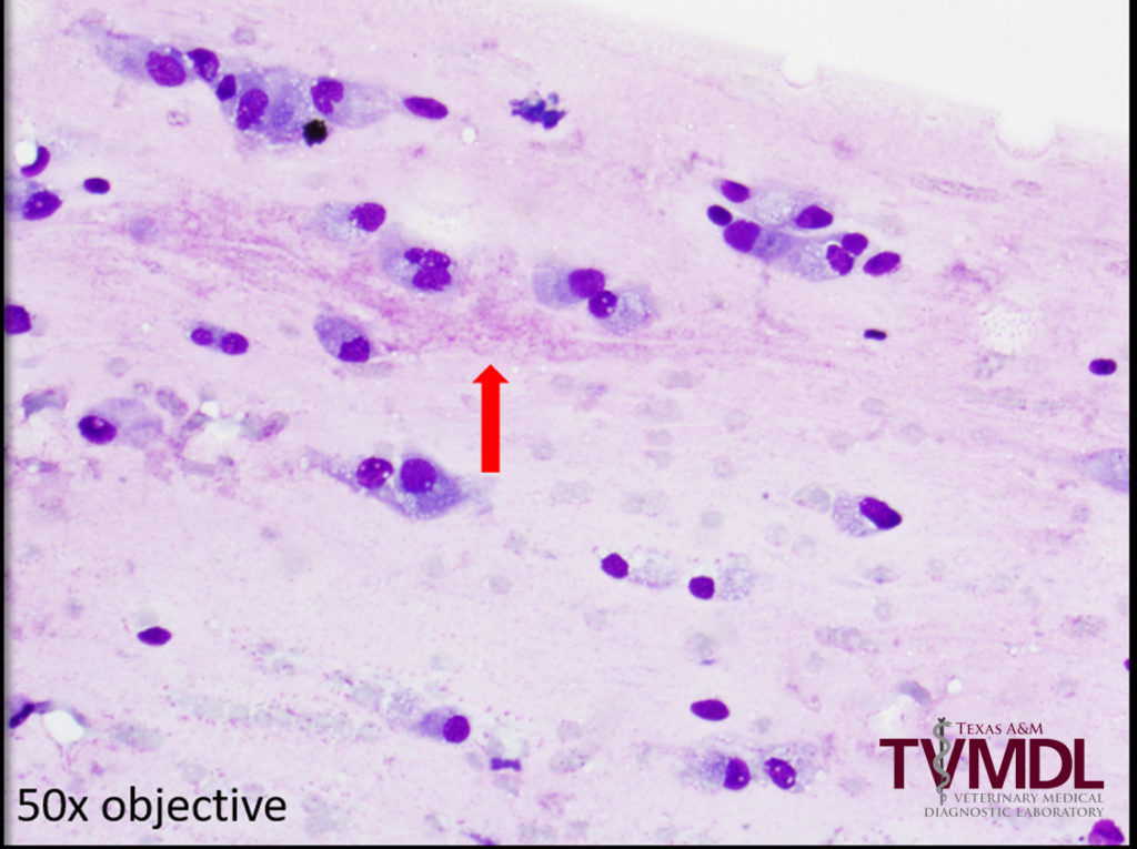
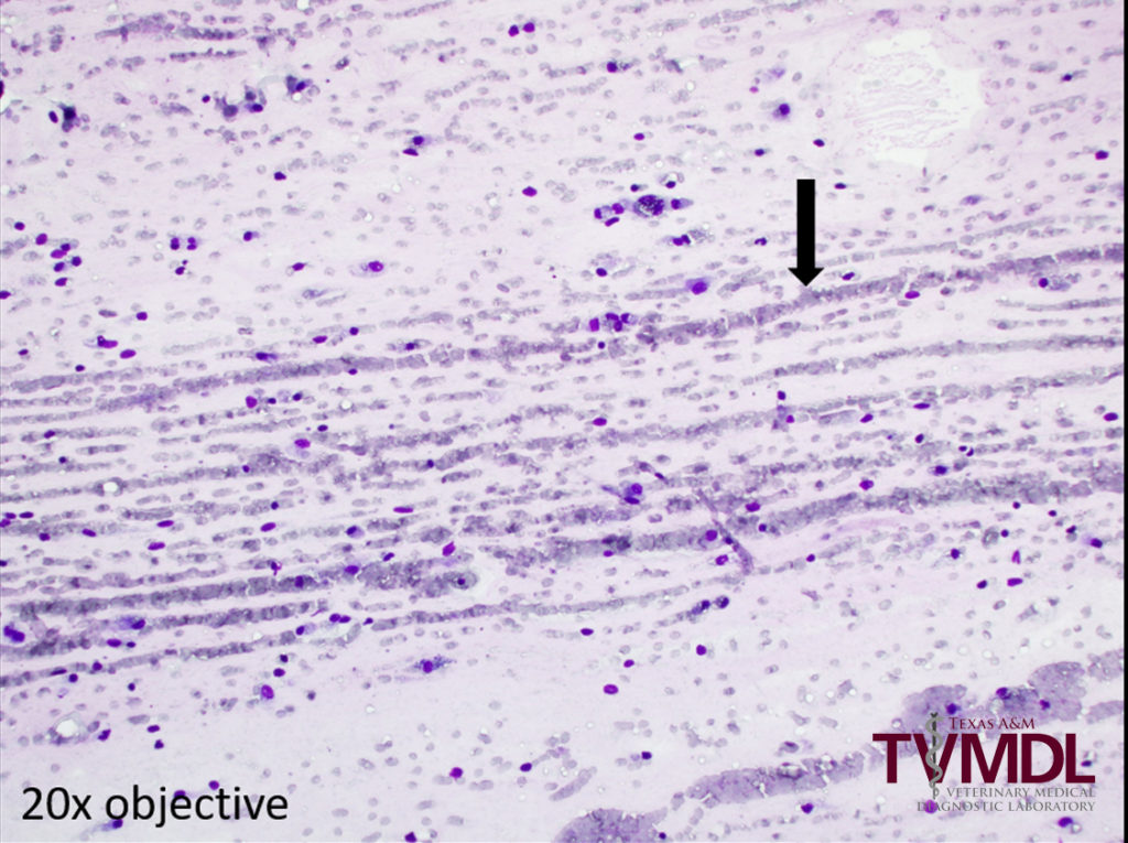
An 11-year-old, spayed female, mixed breed dog was presented to her veterinarian for a mass on the left hind limb. Physical exam revealed a large (~6 cm), irregularly shaped, firm, subcutaneous mass near the left hock. Fine needle aspiration was performed and slides were sent to the Texas A&M Veterinary Medical Diagnostic Laboratory (TVMDL) for evaluation. Cytologic examination revealed a population of neoplastic mesenchymal cells within a dense, myxoid background (Figure 1). Red blood cells and neoplastic cells were often windrowing or arranged in lines (Figure 2) due to myxoid material. The cells displayed mild to moderate anisocytosis (variation in cell size) and anisokaryosis (variation in nuclear size), with some binucleation and prominent nucleoli. The cytologic findings were consistent with aspiration of a sarcoma, and primary consideration was given to a myxosarcoma. Biopsy with histologic examination later confirmed the diagnosis of myxosarcoma.
Myxosarcomas are malignant tumors that are rarely to occasionally observed in veterinary species. The neoplastic cell is a fibroblast; however, the cells produce a dense myxoid matrix, somewhat similar to joint fluid. Cytology can often aid in classification of these lesions; however, biopsy with histologic examination may be needed to differentiate myxoma from myxosarcoma when cell atypia is lacking. Myxosarcomas can be locally invasive, but metastasis is rare. Surgical excision with good margins can be curative.
For more information about this case, contact Dr. Julie Piccione, Clinical Pathology Section Head. To learn more about TVMDL’s testing services, visit tvmdl.tamu.edu or call 1.888.646.5623.

