Canine hepatozoonosis diagnosed via skeletal muscle biopsy
By Erin Edwards, DVM, MS, DACVP
Canine hepatozoonosis is a debilitating, tick-borne disease caused by Hepatozoon americanum, a protozoal parasite transmitted by the Gulf coast tick (Amblyomma maculatum). Affected dogs may have waxing and waning clinical signs and typically present with a fever, weakness, and reluctance to move. Other clinical signs can include muscle atrophy, weight loss, and mucopurulent ocular discharge. Late last year, this disease was diagnosed at the Texas A&M Veterinary Medical Diagnostic Laboratory (TVMDL) in a muscle biopsy from a 3-year-old dachshund-mix dog. The patient was reported to have non-specific clinical signs with a neutrophilic leukocytosis and a WBC of 50,000. The submitting veterinarian sent formalin-fixed punch biopsies from the triceps and semitendinosus muscles to TVMDL. Histopathology revealed a mild granulomatous myositis with scattered onion-skin cysts characteristic of H. americanum.
The geographical distribution of canine hepatozoonosis reflects the distribution of the tick vector, occurring all along the Gulf coast with most cases occurring in Texas, Oklahoma, and Louisiana. Unlike most tick-borne diseases, dogs are infected with H. americanum following ingestion of the tick vector rather than following a tick bite. Infected dogs typically have a severe leukocytosis characterized by a mature neutrophilia, sometimes with a non-regenerative anemia. In many cases, this parasite can induce proliferation of periosteal bony surfaces. These lesions are easily seen on radiographs and resemble those of hypertrophic osteopathy. In hepatozoonosis though, the proximal long bones are more often affected than the distal bones.
A blood smear should be examined in every suspect case, especially those with a compatible leukogram, since in some cases H. americanum gamonts can be found within leukocytes. H. americanum gamonts are typically very rare and confirmative diagnosis often requires submission of skeletal muscle biopsy. These parasites are often widely distributed throughout all skeletal muscle. Since the organisms can be scattered, submission of multiple biopsy specimens is recommended to increase the chance of collecting a diagnostic sample. Histologically, organisms can be found in skeletal and cardiac muscle and may be accompanied by pyogranulomatous inflammation. The parasites are often surrounded by multiple layers of mucopolysaccharide, creating a characteristic “onion-skin” cyst.
For more information on this case, contact Dr. Erin Edwards, veterinary pathologist at the College Station laboratory.
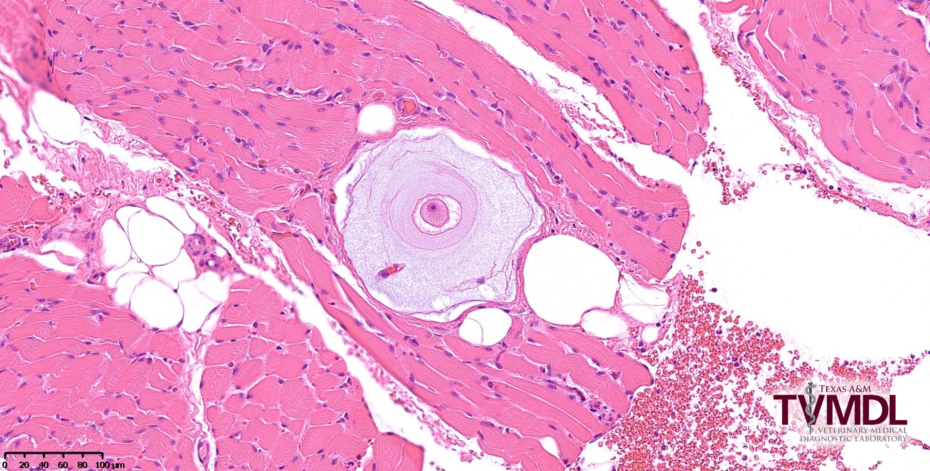
Figure 1: This photomicrograph shows an H. americanum onion-skin cyst within a section of skeletal muscle.