Unusual Manifestation of Papular Stomatitis in a Feedlot Steer
Gayman Helman, DVM, PhD, MA, Will P. Sims, DVM, MS, Gustavo Delhon, PhD
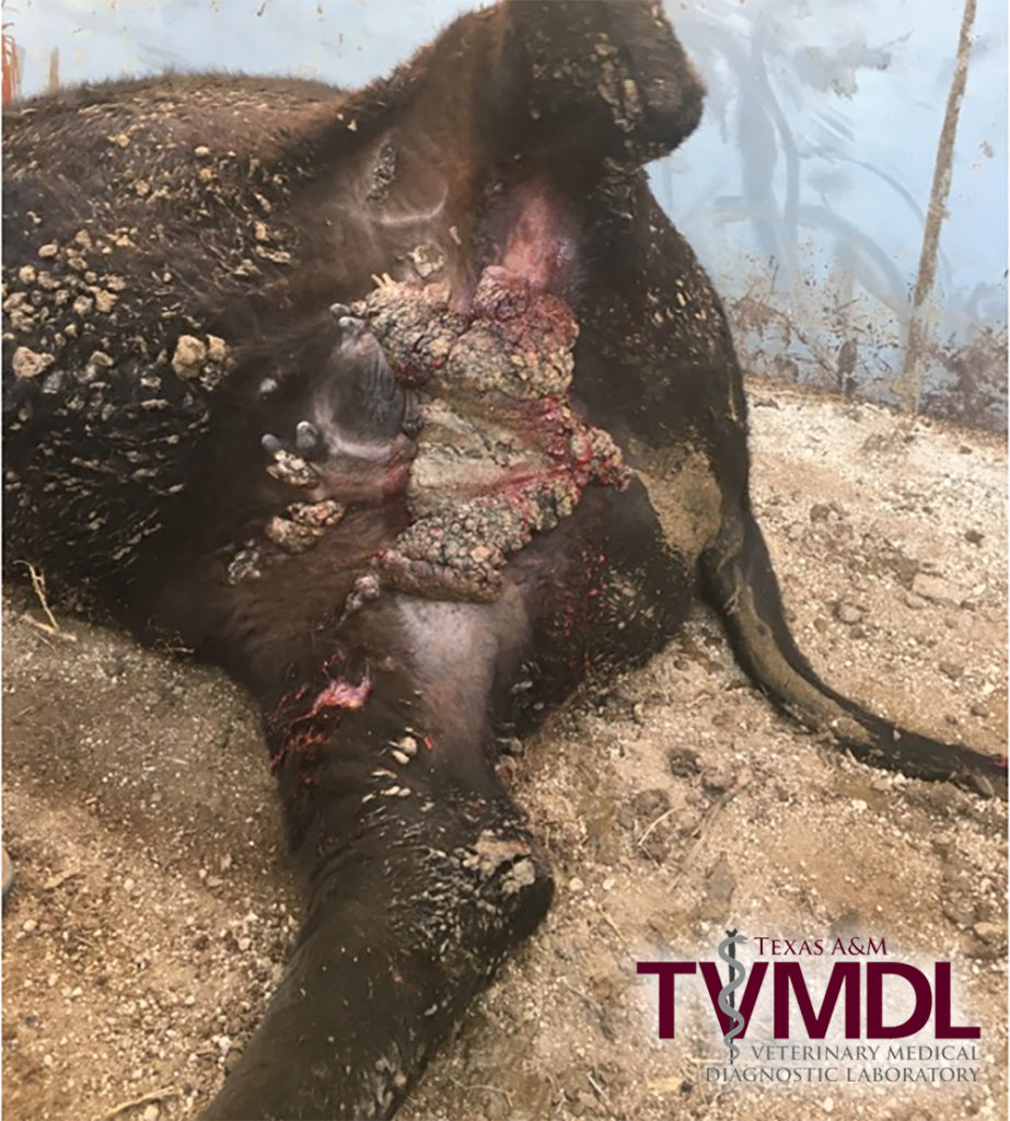
A male-castrated, crossbred steer was noted to have a severe and locally extensive skin disease involving the perineal and inguinal areas with other scattered individual nodular skin masses on other portions of the body (Figure 1). The animal was otherwise healthy and displayed no discomfort from the skin lesions.
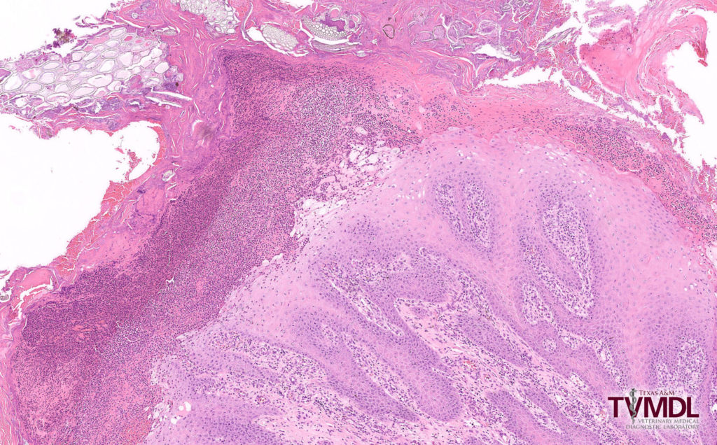
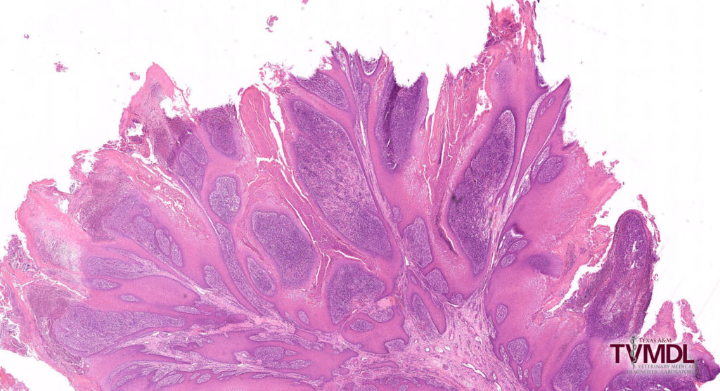
Fresh and fixed skin lesions were submitted to the Texas A&M Veterinary Medical Diagnostic Laboratory (TVMDL) in Amarillo for testing. Grossly, the skin lesions were characterized by marked proliferation of the epidermis with an accumulation of keratin and exudate forming thick crusts on the surface. Microscopically, there was severe epidermal hyperplasia and hyperkeratosis with mixed keratin-purulent crusts adherent to the surface with entrapped foreign material. The underlying dermis was fibrotic with intense infiltrates of lymphoid cells, neutrophils and macrophages (Figures 2 and 3).
Bacterial cultures revealed E. coliand Bacillus sp. as contaminants. A sample of fresh affected skin was forwarded to Dr. Gustavo Delhon at the School of Veterinary Medicine and Biomedical Sciences at the University of Nebraska in Lincoln for virus isolation. A virus was isolated upon blind first passage and the pattern of cytopathic effect (CPE) in the cell culture was compatible with bovine popular stomatitis virus. PCR analysis of viral nucleic acid was confirmatory for bovine papular stomatitis virus.
Papular stomatitis is usually a mild disease in cattle and occurs worldwide. It is associated with proliferative skin lesions in the absence of systemic clinical disease and is most commonly seen as single or multiple cases in young cattle although outbreaks in dairy cattle are reported. The disease may go unnoticed in individual animals as the disease is self-limiting. However, in dairy cattle, lesions on the udder and teats may be associated with loss of production.
The virus is a member of the Poxviridae family and parapoxvirus genus. Other members of the group are Orf virus in sheep (contagious ecthyma), Pseudocowpox virus, which causes “milker’s nodules”, and parapox virus of red deer in New Zealand.
Typical lesions are single to multiple papules on the lips, nares, and oral cavity. Rarely, lesions may appear along the esophagus and in the forestomaches. Lesions usually heal without complications in 4 to 7 days, although there may be residual discoloration of lesion sites for several weeks.
Interestingly, papular stomatitis is a zoonotic disease. Skin lesions have occurred in people having contact with lesions in diseased animals or fomites used in the care and treatment of diseased animals. Lesions typically occur on the hands and severity of disease is variable from mild to severe. Therefore, caution is warranted in handling animals with lesions suggesting papular stomatitis. Appropriate personal protective equipment (PPE) in the form of gloves is warranted.
Immunity to the infection seems to be minimal. Repeat infections are possible. There is no vaccine for bovine papular stomatitis.
The skin lesions in this case were severe and extensive. There are anecdotal reports of individual animals with severe, atypical lesions associated with this virus. One published report of atypical lesions was associated with coinfection with multiple strains of the virus 1. The etiology of such atypical severe cases is unknown, but perhaps may be related to immunocompromise.
For more information on this case, contact Amarillo Resident Director Dr. Gayman Helman or Dr. Will Sims, veterinary pathologist at TVMDL-Amarillo. To learn more about TVMDL’s test offerings, visit tvmdl.tamu.edu or call TVMDL-Amarillo at 1.888.646.5624 or TVMDL-College Station at 1.888.646.5623.

Figure 1. Extensive portions of the skin disease were localized to the perineal and inguinal areas. Individual nodular masses were scattered on other parts of the body.

Figure 3. The underlying dermis was fibrotic and also as having intense infiltrates of lymphoid cells, neutrophils, and macrophages.
References:
- Archives of Virology, 2015, 160(6): 1527-32.
- Fenner’s Veterinary Virology, NJ MacLachlan and EJ Dubovi editors, 5thedition, 2017, pp 47-78, 157-174.
