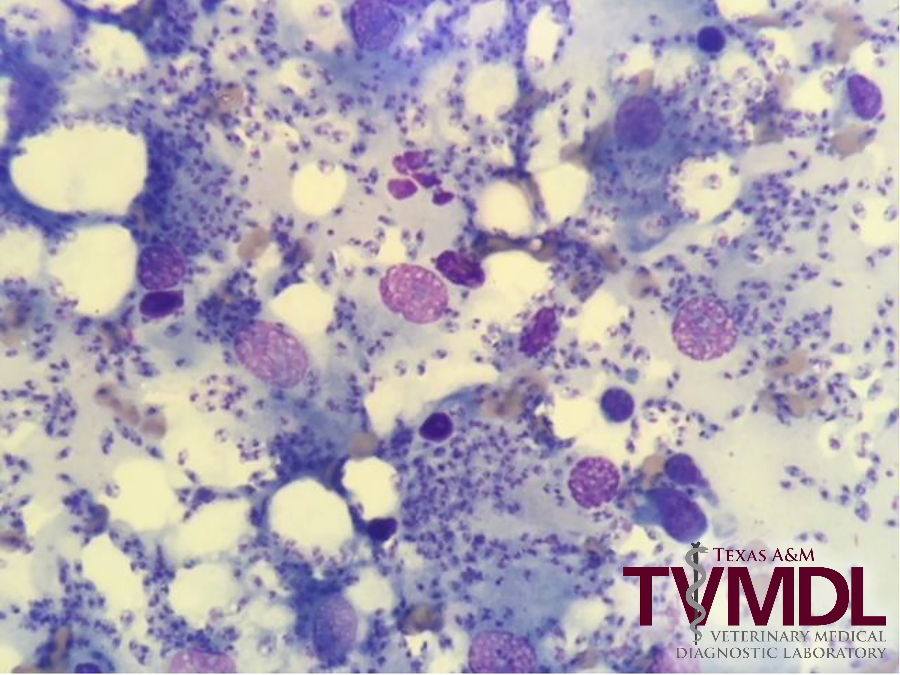
Leishmaniasis diagnosed in 9-year-old cat
By Judith Akins, DVM, MS
A fine needle aspirate of a mass on the left ear pinnae of a 9-year-old, spayed, female domestic shorthaired cat was submitted to the Texas A&M Veterinary Medical Diagnostic Laboratory (TVMDL) cytology service. History stated the lesion began as a small scab about a month before the sample collection, but had grown rapidly to its present size of 1.5 cm. The cat was not reported to have any other clinical signs.
The examined slides were cellular with large numbers of macrophages. The macrophages were actively phagocytizing large numbers of protozoal amastigotes. The amastigotes were small (1.5 to 5 microns) and round. The centers contained a dark staining nucleus with a characteristic rod shaped kinetoplast. The diagnosis was granulomatous inflammation with protozoa consistent with Leishmania spp.
Leishmaniasis is an infectious disease caused by a protozoa of the genus Leishmania. This protozoon is typically found in temperate and tropical climates. Reservoirs for the protozoa differ depending upon the location in the world (New World vs. Old World). For New World leishmaniasis, the endemic region includes South and Central America, Mexico, and pockets of the United States. The disease is transmitted by sandfly vectors from an infected to an uninfected host. There have been reports in the United States of infections in dogs, cats, and humans with the following species: L. mexicana complex, L. braziliensis complex, and L. donovani complex. Treatment for this disease in animals is difficult and may be prolonged.
To learn more about this case, contact Dr. Judith Akins, clinical pathologist. For more information about TVMDL’s test offerings, visit tvmdl.tamu.edu or call 1.888.646.5623.
Reference
Veterinary Pathology, Vol.47, Issue 6, 2010. Trainor, et. al.
