Isolation of Nannizziopsis sp. from skin lesions of a veiled chameleon (Chamaeleo calyptratus)
By Owais Khan, BVSc & AH, MVSc, Will Sims, DVM, MS, and Amy Swinford, DVM, MS
An eight-month-old veiled chameleon exhibited white, patchy, crusty skin lesions on the dorsal crest above the hind legs (Fig. 1). A skin biopsy specimen was submitted to the Texas A&M Veterinary Medical Diagnostic Laboratory (TVMDL) for histopathology and a swab and skin crust samples were submitted for routine bacterial and fungal culture.
The swab sample for bacterial culture was inoculated onto Blood agar and Tergitol agar plates which were incubated at 37oC and skin crusts were inoculated onto Sabouraud Dextrose Agar (SDA) and Dermatophytes Test Medium (DTM) agar plates and were incubated in an ambient environment at 22oC.
No bacteria were isolated from the swab sample. Following five days of incubation, five individual pure white colonies on the SDA and one individual colony on the DTM plate were isolated (Fig.2). The reverse side of the colonies were initially yellow (Fig. 3), but over time changed to cream-colored, and the colonies became chalky in consistency. The microscopic morphology was similar to Chyrysosporium species (Fig.4) and final identification was done by the sequencing of the internal transcribed spacer region of 18s ribosomal RNA. The fungal isolate had 98% homology to Nannizziopsis draconii strain CBS 133987 referenced in GenBank. Microscopically, the biopsy sample showed the presence of fungal hyphae within the epidermis, with mild amounts of inflammation demonstrated with the GMS stain (Fig.5). It is possible this is the first report of isolation of Nannizziopsis draconii from a veiled chameleon.
Veiled chameleons are one of the most widely available types of chameleon and are commonly kept as pets or in private collections. The veiled chameleon is also known as Yemen Chameleon, as it is native to the Arabian Peninsula. Fungal skin infections (dermatomycoses) are common in reptiles and have been reported in snakes, bearded dragons, iguanas, chameleons, and lizards. A fungus identified as Chrysosporium anamorph of Nannizziopsis vriesii (CANV) as well as other Chrysosporium species have emerged as leading causes of fungal dermatitis in reptiles. The natural habitat of these keratinophilic fungi are environment (soil, cage substrates etc.), and damage to skin, poor husbandry and stress factors predispose captive reptiles to the fungal infection.
For more information about this case, contact Dr. Owais Khan, TVMDL Amarillo bacteriology section head, Dr. Will Sims, veterinary pathologist at TVMDL Amarillo, or Dr. Amy Swinford, associate director at the College Station laboratory. To learn about TVMDL’s test offerings, visit tvmdl.tamu.edu or call TVMDL Amarillo at 1.888.646.5624 or TVMDL College Station at 1.888.646.5623.
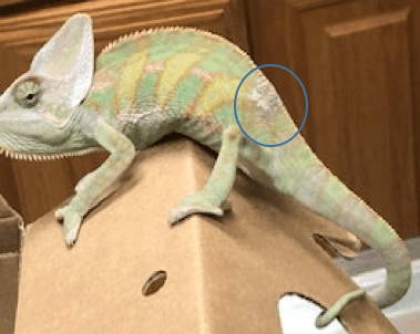
Figure 1. Circled area showing white crust
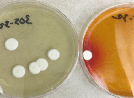
Figure 2. Five day old culture on SDA and DTM (fornt side)
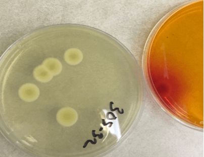
Figure 3. Five day old culture in SDA and DTM (reverse side)
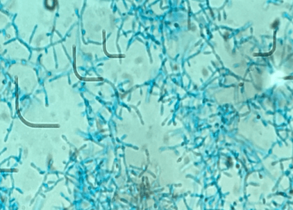
Figure 4. Microscopic appearance of the isolate showing aleuriconidia borne on hyphae (LPCB, X400)
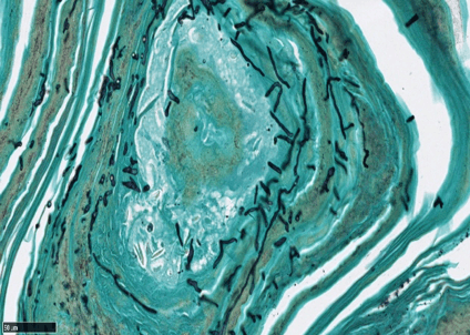
Figure 5. Non branching fungal hyphae in epidermal keratin layer (GMS, X400)