Pulmonary Toxoplasmosis Diagnosed in a Young Cat
By Erin Edwards, DVM, MS, DACVP
Pulmonary toxoplasmosis was recently diagnosed in a stray, male tabby cat approximately 1-year-old. This cat was brought to a local veterinary clinic for lethargy and respiratory distress and died within 24 hours of treatment. A pneumonia was identified at necropsy and formalin-fixed samples of lung were sent to the Texas A&M Veterinary Medical Diagnostic Laboratory (TVMDL) for histopathology. Examination of the slides revealed a severe, necrotizing, interstitial pneumonia with numerous intralesional protozoal tissue cysts consistent with Toxoplasma gondii (Figure 1).
Cats are the only recognized definitive host for toxoplasmosis, and outdoor hunting cats are the most likely to be infected. Once infected, cats will shed heavy amounts of oocysts for a few weeks which will then begin to taper. Oocysts sporulate after 1-5 days at which point they become infective to other mammals and birds that ingest them. Once ingested by another host, the parasites typically form multiple tissue cysts in the skeletal muscle and organs, and these cysts generally do not cause any harm. In immunocompromised or susceptible hosts, though, these cysts can begin to rapidly divide, causing tissue necrosis.
Cats typically do not have clinical disease from toxoplasmosis. Infection with FIV or FeLV is known to cause immunosuppression, predisposing cats to disease from toxoplasmosis. Similarly, dogs typically only have toxoplasmosis when there is an underlying cause of immune suppression, most often co-infection with canine distemper virus. Affected dogs and cats typically have systemic infection, with the lungs, brain, heart, and liver being the most commonly affected organs. Certain species of animals are highly susceptible to toxoplasmosis without having another cause of immunosuppression, particularly kangaroos, wallabies, lemurs, and New World primates. In small ruminants, toxoplasmosis causes late term abortion and the pathologic lesion is characterized by cotyledonary necrosis.
Of note, toxoplasmosis is a disease of public health interest as humans can be infected when exposed to this parasite. Pregnant women and HIV-infected individuals are at greatest risk and infection can cause fetal abnormalities or miscarriage. Although exposure can occur through contact with feces while scooping a litter box, this route of exposure is far less common than other routes including consumption of unwashed vegetables that were grown in contaminated soil or consumption of under-cooked infected meats. To prevent disease and avoid exposure, the Center for Disease Control (CDC) recommends to thoroughly wash and cook all produce and meats, to scoop the litterbox within 24 hours of fecal deposition (before sporulation occurs), or, if possible, to have another member of the household scoop the litterbox during pregnancy.
For more information about this case, contact Dr. Erin Edwards, veterinary pathologist. To learn more about TVMDL’s testing services, call 1.888.646.5623 or visit tvmdl.tamu.edu.
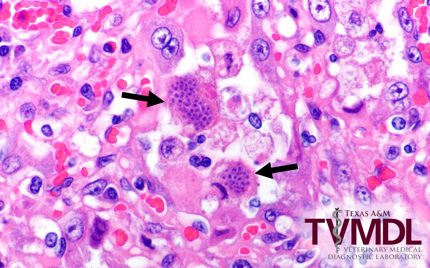
Figure1. Photomicrograph depicting Toxoplasma gondii.
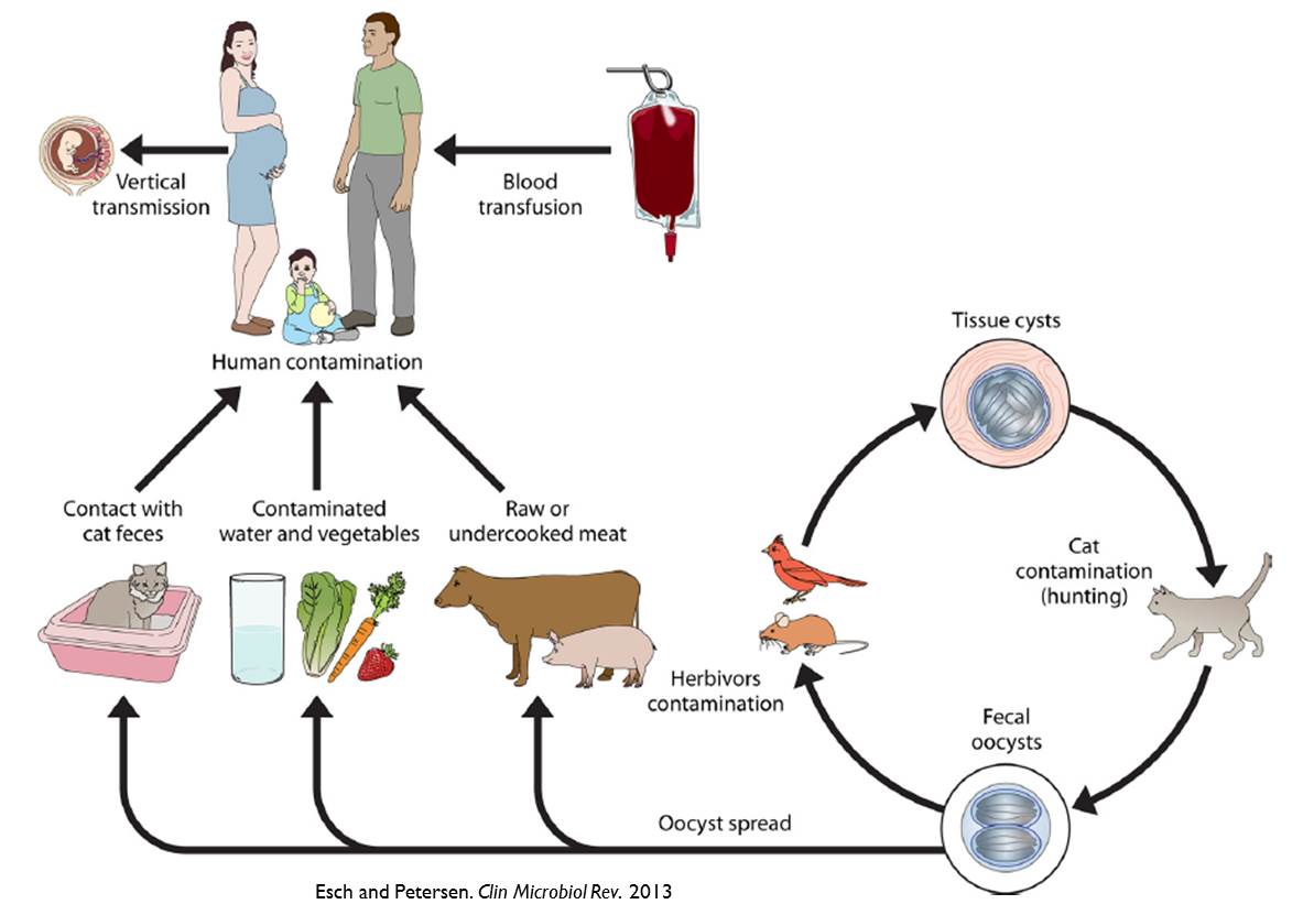
Figure 2. Infection cycle of Toxoplasma gondii.