Visceral Mast Cell Disease In A Cat
By Julie Piccione, DVM, MS, DACVP
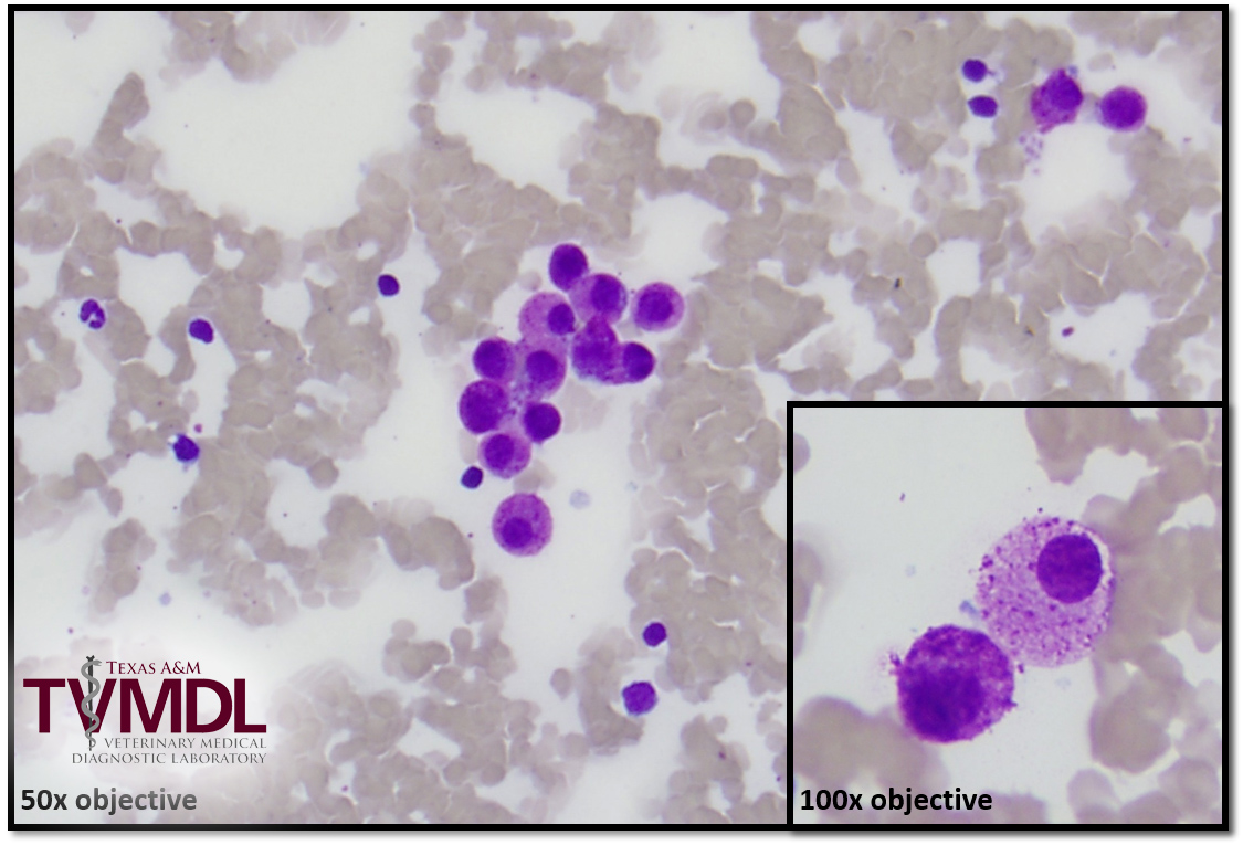
An 11-year-old, female spayed, domestic short haired cat was presented to her veterinarian for increased appetite, weight loss, and occasional vomiting. Besides a low body condition score, no overt abnormalities were identified on physical exam. A minimum database (CBC, chemistry, T4, and urinalysis) was performed at the Texas A&M Veterinary Medical Diagnostic Laboratory (TVMDL). The only abnormalities detected were marked poikilocytosis (irregularly shaped RBCs) and a mild leukopenia (see figure 1). An abdominal ultrasound was performed at Texas A&M College of Veterinary Medicine and Biomedical Sciences (TAMU CVM) and revealed mild splenomegaly with a mottled echotexture. A fine needle aspirate of the spleen revealed numerous well-granulated, uniform mast cells, and a diagnosis of splenic mastocytosis was made (see figure 2).

Figure 1: Peripheral blood smear in a cat. Note the irregularly shaped red blood cells. Wright-Giemsa stain.

Figure 2: Fine needle aspiration cytology of the spleen. Mast cells are significantly increased. Wright-Giemsa stain.
Splenic mast cell disease in cats can include cutaneous and visceral forms. Visceral mast cell disease can include gastrointestinal and splenic neoplasia, also known as splenic mastocytosis. Clinical signs of visceral mast cell disease in cats often include vomiting, diarrhea, weight loss, and weakness. Gastrointestinal mast cell disease is often associated with a poor prognosis. However, cats with splenic mastocytosis can have long survival times with splenectomy, when distant metastases are absent. Poikilocytosis and leukopenia can be associated with splenic disease, as seen in this patient.
To learn more about this case, contact Dr. Julie Piccione, clinical pathology section head at the College Station laboratory. For more information on TVMDL’s test catalog, visit tvmdl.tamu.edu or call 1.888.646.5623.