• Skip to primary navigation
  • Skip to main content
  • Skip to primary sidebar
  • Skip to footer
  • Locations:
  • Canyon
  • Center
  • College Station
  • Gonzales
  • TVMDL Career Center
  • Contact Us

Texas A&M Veterinary Medical Diagnostic Laboratory

Apple App

download on the Apple App Store
download the app on Google Play

Search

Translate:

  • Find a Form
  • Deliver a Sample
    • Specimen Collection Information
    • Packaging Samples for Shipment
    • Drop Off a Sample
    • Cremation
    • Order Supplies
  • Client Portal
    • Get Test Results
    • SVA Portal
    • Pay a Bill
  • Become a Client
  • Resources
    • TVMDL Bovine Testing Guidance and Reference Material
    • Diagnostic Plans
      • Bovine Syndromic Diagnostic Plans
      • Equine Syndromic Diagnostic Plans
      • Small Ruminant Syndromic Diagnostic Plans
      • Small Animal Syndromic Diagnostic Plans
    • Education Library
    • Case Study Library
    • Electronic Reporting: QR Coding Process
    • CoreOne Resources
  • About Us
    • Locations and Hours
    • Agency Leadership
    • Speaker’s Bureau
    • Texas Pullorum-Typhoid Program
    • TVMDL Mobile app
    • Contact Us
  • Locations
    • College Station Laboratory
    • Canyon Laboratory
    • Center Laboratory
    • Gonzales Laboratory
  • Contact Us

Histomoniasis “Blackhead” in Bobwhite Quail

June 25, 2018 by Mallory Pfeifer

Histomoniasis “Blackhead” in Bobwhite Quail
By Gabriel Senties-Cue, MVZ, EPAA, MS

Histomoniasis, a protozoal disease of birds also known as “Blackhead”, was diagnosed in a flock of Bobwhite quail. Three dead females and one dead male, 8-month-old, Bobwhite quail were submitted to the Texas A&M Veterinary Medical Diagnostic Laboratory (TVMDL) with a clinical history of increased mortality. History indicated the flock exhibited enteritis and had been dewormed the previous month. Upon necropsy examination, multifocal, pale and irregular in size foci were seen scattered over the surface of the liver. Both ceca were dilated with yellowish-tan caseous cores in the lumen and small raised nodules on the mucosa. Histologically, large numbers of protozoal organisms consistent with Histomonas meleagridis associated with inflammatory cells were seen in the submucosa and muscular layer of ceca (Fig. 1). In liver sections extensive areas of necrosis with vacuoles, some containing protozoal organisms undergoing degeneration, were seen. The lack of a treatment for histomoniasis has caused this disease to become common again in gallinaceous species.

For more information about this case, contact Dr. Senties-Cue, resident director at TVMDL Center. To learn more about TVMDL’s test catalog, visit tvmdl.tamu.edu or call 1.888.646.5623.


Photomicrograph depicting Histomonas meleagridis

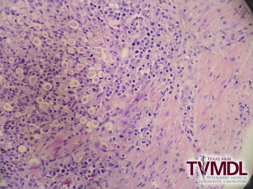
Photomicrograph depicting Histomonas meleagridis.

Filed Under: Case Study Tagged With: Avian, bobwhite quail, center, TVMDL

Primary Sidebar

Latest Case Studies

  • Neurologic Disease due to Bovine Herpesvirus-5 (BHV-5) Infection in a 3-week-old Charolais mix calf

    February 27, 2023

  • Rare case of ocular onchocerciasis in a dog from south Texas

    January 25, 2023

  • Cerebrospinal Nematodiasis (Visceral Larval Migrans) in Birds

    September 13, 2022

  • Spironucleosis (Hexamitiasis) in Quail

    August 9, 2022

  • Endocarditis in a White-Tailed Deer Caused by Trueperella pyogenes

    August 9, 2022

Footer

For Employees

  • Employee Email
  • TVMDL Rules & Procedures
  • TVMDL Career Center
  • Emergency Alert Systems
  • AgriLife People Directory
  • AgriLife Administrative Services
  • TAMUS Single Sign On
  • Facebook

State of Texas

  • Texas.gov Portal
  • Texas Veterans Portal
  • Statewide Search
  • Texas Homeland Security
  • Risk, Fraud, & Misconduct Hotline

Policies

  • Privacy and Security Policy
  • Accessibility Policy
  • Texas A&M AgriLife
  • Texas A&M Veterinary Medical Diagnostics Laboratory
  • Texas A&M Forest Service
  • Texas A&M AgriLife Extension Service
  • Texas A&M AgriLife Research

483 Agronomy Rd
College Station, TX 77843