Feline Osteosarcoma
By Judith Akins, DVM, MS
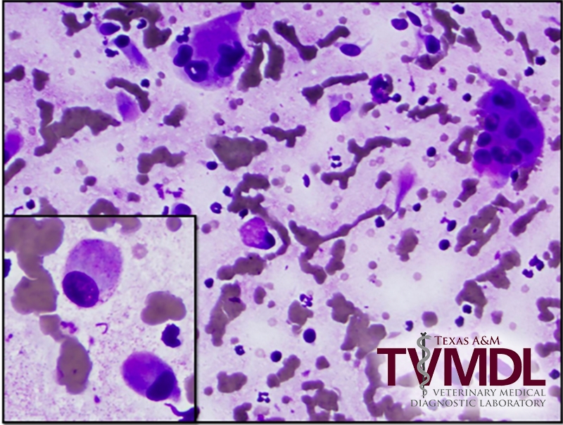
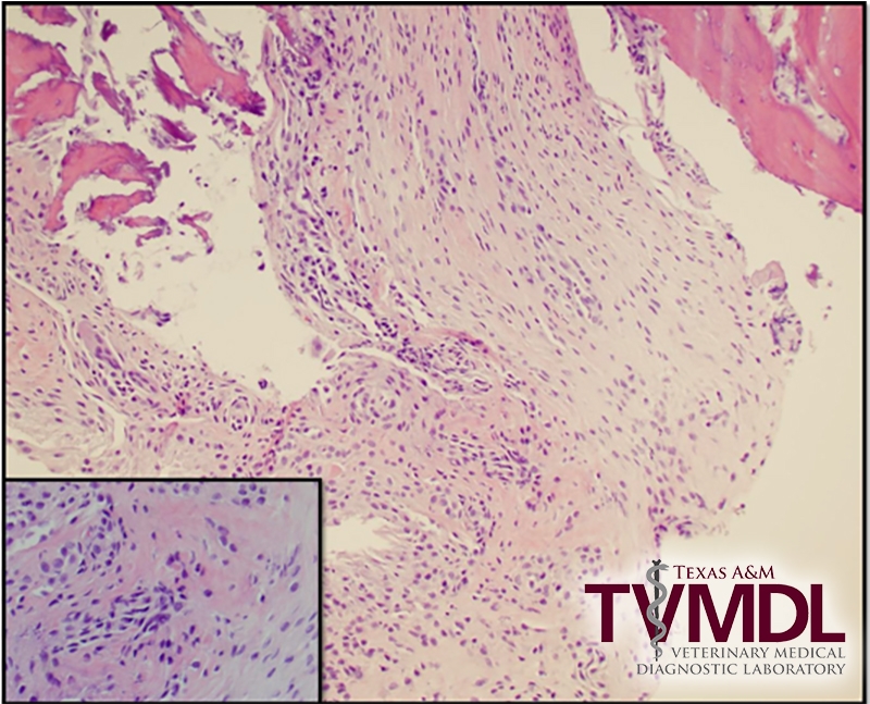
A small piece of carpal bone from an eight-year-old domestic short haired cat (DSH) was submitted to the Texas A&M Veterinary Medical Diagnostic Laboratory (TVMDL) for histopathology. The provided history stated the cat had a lytic and proliferative lesion of the “carpal bones”. An impression smear of the boney lesion was also submitted. The slides for cytology contained large numbers of atypical spindle shaped cells. There were a few scattered multi-nucleated cells (interpreted as osteoclasts). There were no increased numbers of leukocytes or any etiologic agents. The biopsy specimen was small but consisted of sheets of spindle cells that surrounded amorphous pale eosinophilic staining material (interpreted as osteoid). The diagnosis was osteosarcoma.
Osteosarcomas in cats are much less common than in dogs, but account for approximately 70% of the malignant bone tumors in cats. In one study, the age range of affected cats was 8 to 10.5 years of age. The clinical course of the disease process tends to be slower in cats than in dogs. In one study looking at 12 cats, there was a median survival time of 49.2 months following amputation of the limb with the osteosarcoma. Typically, metastasis is less common in cats than in dogs, but if metastasis does occur, the lung is the primary site.
For more information about this case, contact Dr. Judith Akins, Clinical Pathologist at the College Station laboratory. To learn more about TVMDL’s test catalog and services, visit tvmdl.tamu.edu or call 1.888.646.5623.

