Testicular Coccidioidomycosis in a Dog
By Jay Hoffman, DVM, PhD
With over 800,000 tests run annually, TVMDL encounters many challenging cases. Our case study series will highlight these interesting cases to increase awareness among veterinary and diagnostic communities.
Fixed and fresh tissue samples from the testicles of a dog were received at the Texas A&M Veterinary Medical Diagnostic Laboratory (TVMDL) for histopathologic evaluation and bacterial culture. The dog was approximately 2 years old and from south Texas. Both of the submitted testicles were described as being very hard and the dog was described to be very thin.
Histologic evaluation of the testicle revealed dense infiltrates of neutrophils, macrophages, lymphocytes and plasma cells randomly scattered throughout the testicle and surrounding tunic. Occasionally, the inflammatory infiltrate surrounded fungal spherules ranging from approximately 30-60 min diameter. The spherules had thick, double contoured walls and contained numerous endospores ranging from 2-5 min diameter. These fungal spherules were morphologically consistent with Coccidioides immitis. Surrounding the inflammatory exudate were thick strands of fibrous connective tissue. Most of the adjacent seminiferous tubules were collapsed and were lined by Sertoli cells. The adjacent epididymis was devoid of mature spermatozoa.
Coccidioidomycosis is caused by the dimorphic fungus Coccidioides immitis. The disease is most common in the endemic areas of the United States which includes the arid parts of California, Arizona, and Texas. In tissues, distinctive spherule with endospores are observed. In the environment, infective arthroconidia are found. Inhalation of the airborne arthroconidia is the usual route of infection. This organism primarily causes a respiratory infection in human beings, dogs, horses, sheep, cattle, swine, and cats. The majority of respiratory infections are benign and self-limiting; in humans this form is known as San Joaquin Valley fever. A small percentage of the infections disseminate from the lungs and cause generalized disease. Among the domestic species, the disseminated form of the disease is most common in dogs with Boxers and Doberman pinschers being overrepresented. When disseminated, the most common sites affected include the bone, heart, lymph nodes, skin, heart, brain, eyes, liver, spleen, kidney, and testes. In some cases, there is widespread dissemination of the organism without observable pulmonary lesions. This is thought to be due to resolution of the lung lesion after dissemination. It is thought that the testicular lesion in this animal was the result of a disseminated infection. The poor body condition described in the clinical history supports this suspicion.
Definitive diagnosis of coccidioidomycosis is by identification of the characteristic fungal spherules in tissue sections, although the spherules are typically present in low numbers. Serum antibodies to Coccidioides immitiscan be helpful in the acute stages of the disease with high titers indicative of infection.
Contact Pathology Branch Chief Dr. Jay Hoffman for questions about this case. For more information about TVMDL’s test offerings, visit tvmdl.tamu.edu or call 1.888.646.5623.
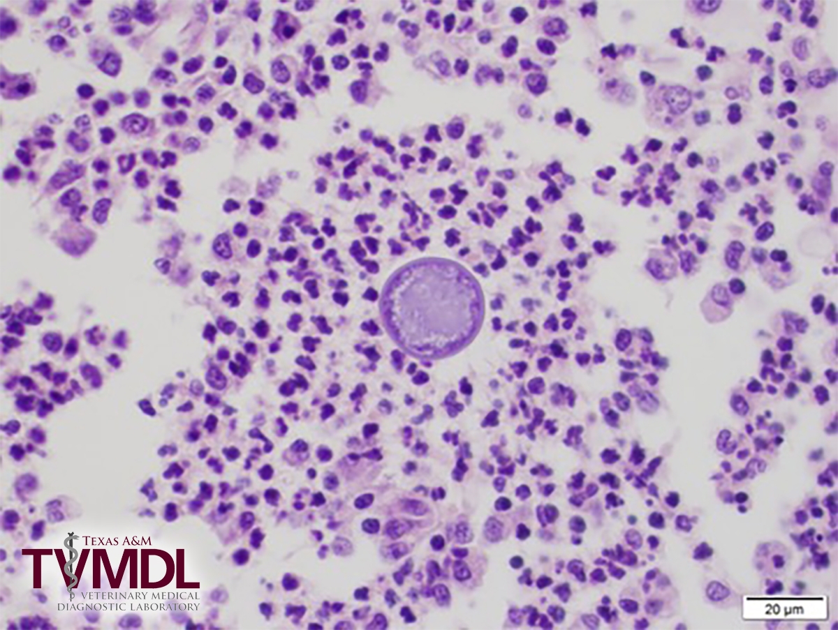
Testicular tunic from dog. Characteristic fungal spherule of Coccidioides immitis with pyogranulomatous inflammation.