Inclusion body hepatitis (IBH) in broiler chickens
By Gabriel Senties-Cue, MVZ, EPAA, MS
With over 800,000 tests run annually, TVMDL encounters many challenging cases. Our case study series will highlight these interesting cases to increase awareness among veterinary and diagnostic communities.
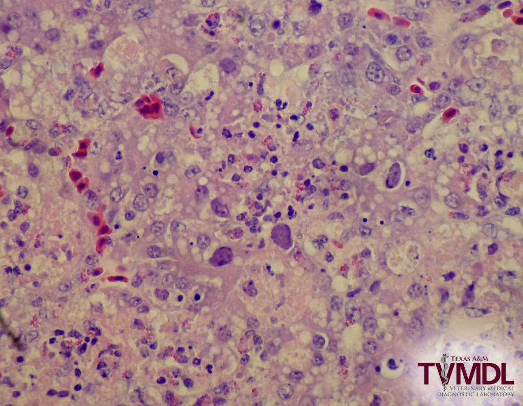
Inclusion body hepatitis (IBH) was diagnosed in a flock of 12-day- old, meat type chickens. Six live and 4 dead birds were submitted to the TVMDL-Center Lab with a history of increased mortality, swollen kidneys, and whitish spots livers. At the necropsy examination livers were pale, mottled with a reticular pattern and with subcapsular hemorrhages; heart sacs had accumulation of a clear fluid in the pericardium; bursas of Fabricius were atrophied; kidneys were swollen and pale; and in pancreases there were pale pin-point spots. The histopathological examination of liver and pancreas sections revealed multifocal to coalescent areas of necrosis associated with mixed inflammatory cell infiltration and large, basophilic intranuclear inclusions, which are characteristic of IBH. IBH is caused by Group I avian adenoviruses, which are transmitted vertically and horizontally. IBH in chickens is commonly associated with immunosuppression.

Large basophilic intranuclear inclusions in hepatocytes associated with necrosis, vacuolation and with mixed inflammation.
For more information on this case, contact Dr. Gabriel Senties-Cue, Resident Director at Center laboratory. To learn more about TVMDL’s test catalog, visit tvmdl.tamu.edu or call 1.888.646.5623.
Visit TVMDL’s case study library to read this case and others.