Equine Protozoal Myeloencephalitis (EPM) Diagnosed in a Horse
By Erin Edwards, DVM, MS, DACVP
With over 800,000 tests run annually, TVMDL encounters many challenging cases. Our case study series will highlight these interesting cases to increase awareness among veterinary and diagnostic communities.
Recently, the brain of a 7-year-old Quarter horse mare was sent to the Texas A&M Veterinary Medical Diagnostic Laboratory (TVMDL) for histopathology. This horse was reported to have clinical neurological signs. Prior to histologic examination, the brain was determined to be rabies-negative by the Texas Department of State Health Services. Histologically, vessels in the brain and brainstem were multifocally surrounded by small cuffs of lymphocytes and plasma cells. These tissues also contained scattered foci of lymphohistiocytic inflammation with rare small areas of necrosis (Fig. 1A). Intermingled with these inflammatory cells there were a few protozoal schizonts, prompting the diagnosis of Equine Protozoal Myeloencephalitis (EPM) for this horse (Fig. 1B).

The most common cause of EPM is Sarcocystis neurona, a protozoal parasite that is widespread across the United States. The Virginia opossum (Didelphis virginiana) is the definitive host for this parasite, and horses are typically exposed after ingesting feed, water, or other sources contaminated with the feces of infected opossums. Not all exposed horses develop clinical disease though, and factors that elicit the spread of the parasite to the central nervous system are poorly characterized. The disease is more prevalent during late summer and fall, but this case demonstrates that EPM can occur outside of those seasons, at least in Texas. 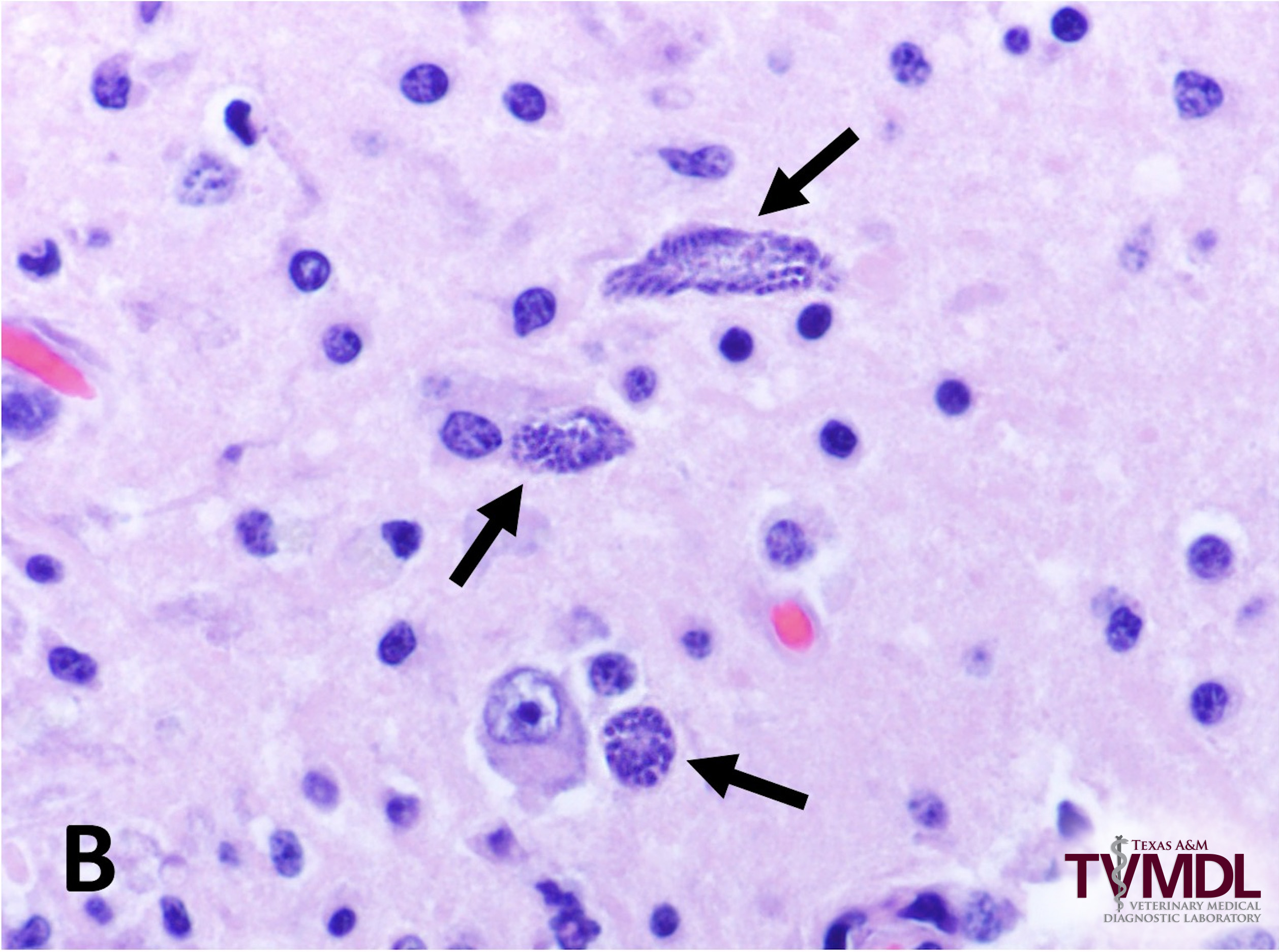
Affected horses will often show clinical evidence of neurologic disease. The parasite can affect any part of the central nervous system. Signs can vary widely, but some common clinical signs include ataxia, weakness, muscle atrophy, tremors, facial paralysis, and recumbency.
There are many options for diagnosing EPM. Antemortem, a serum titer test can be performed to search for antibodies to Sarcocystis neurona in horses suspected of having EPM. Detection of antibodies though, only confirms exposure to the parasite and will not confirm EPM as the cause of associated neurologic disease. Titers performed on cerebrospinal fluid (CSF) are more suggestive of active disease. A polymerase chain reaction test (PCR) for Sarcocystis neurona can also be performed on CSF. This test is typically not favored due to a low sensitivity and high numbers of false negatives. Postmortem, histopathology is often diagnostic. In some cases, organisms may be low in number and difficult to find. In these cases, other histologic lesions are often suggestive of the disease, though additional testing such as immunohistochemistry or PCR may be necessary to confirm the diagnosis. Rare cases of EPM are also caused by Neospora hughesi, which cannot be reliably differentiated histologically from Sarcocystis neurona. Immunohistochemistry and/or PCR are needed for ultimate differentiation.
Besides EPM, there are many other differentials for neurologic disease in horses. One disease that should always be ruled out is rabies. A rabies test should be performed on any horse that died from or was euthanized for neurologic disease. For the safety of any exposed individuals and laboratory personnel handling samples in the lab, TVMDL policy requires negative rabies test results from the Texas Department of State Health Services (TDSHS) before additional tests are performed on tissues from rabies suspect cases. Rabies samples can be submitted directly to the TDSHS or can be forwarded from TVMDL.
For more information about this case, contact Dr. Edwards, Veterinary Pathologist at the College Station laboratory. To learn more about TVMDL’s test catalog, visit tvmdl.tamu.edu or call 1.888.646.5623.