• Skip to primary navigation
  • Skip to main content
  • Skip to primary sidebar
  • Skip to footer
  • Locations:
  • Canyon
  • Center
  • College Station
  • Gonzales
  • TVMDL Career Center
  • Contact Us

Texas A&M Veterinary Medical Diagnostic Laboratory

Apple App

download on the Apple App Store
download the app on Google Play

Search

Translate:

  • Find a Form
  • Deliver a Sample
    • Specimen Collection Information
    • Packaging Samples for Shipment
    • Drop Off a Sample
    • Cremation
    • Order Supplies
  • Client Portal
    • Get Test Results
    • SVA Portal
    • Pay a Bill
  • Become a Client
  • Resources
    • TVMDL Bovine Testing Guidance and Reference Material
    • Diagnostic Plans
      • Bovine Syndromic Diagnostic Plans
      • Equine Syndromic Diagnostic Plans
      • Small Ruminant Syndromic Diagnostic Plans
      • Small Animal Syndromic Diagnostic Plans
    • Education Library
    • Case Study Library
    • Electronic Reporting: QR Coding Process
    • CoreOne Resources
  • About Us
    • Locations and Hours
    • Agency Leadership
    • Speaker’s Bureau
    • Texas Pullorum-Typhoid Program
    • TVMDL Mobile app
    • Contact Us
  • Locations
    • College Station Laboratory
    • Canyon Laboratory
    • Center Laboratory
    • Gonzales Laboratory
  • Contact Us

Necrotic enteritis in broilers

March 2, 2018 by Mallory Pfeifer

Necrotic enteritis in broilers
By Gabriel Senties-Cue MVZ, EPAA, MS

With over 800,000 tests run annually, TVMDL encounters many challenging cases. Our case study series will highlight these interesting cases to increase awareness among veterinary and diagnostic communities.

Necrotic enteritis associated with coccidiosis was diagnosed in 25-day-old broilers. Six live broilers with a history of increased mortality with birds dying with their legs straight out were submitted for laboratory examination. At necropsy, the mucosa of the jejunum and ileum had a “cloth towel” appearance with loose, small mucosal flakes. Moderate numbers of coccidia were detected by the wet mount technique. The diagnosis of necrotic enteritis was confirmed by histopathology, which revealed necrosis of the upper part of the intestinal mucosa associated with large numbers of both rod-shaped bacteria and coccidia. Necrotic enteritis is caused by toxins secreted by Clostridium perfringens, which proliferates in the intestinal lumen when damage of the intestinal mucosa occurs. Coccidiosis is the main predisposing factor to necrotic enteritis.

To learn more about this case, contact Dr. Senties-Cue, Center Resident Director. For more information on TVMDL’s test catalog, visit tvmdl.tamu.edu.

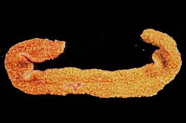
Intestinal mucosa exhibiting a “cloth towel” appearance.

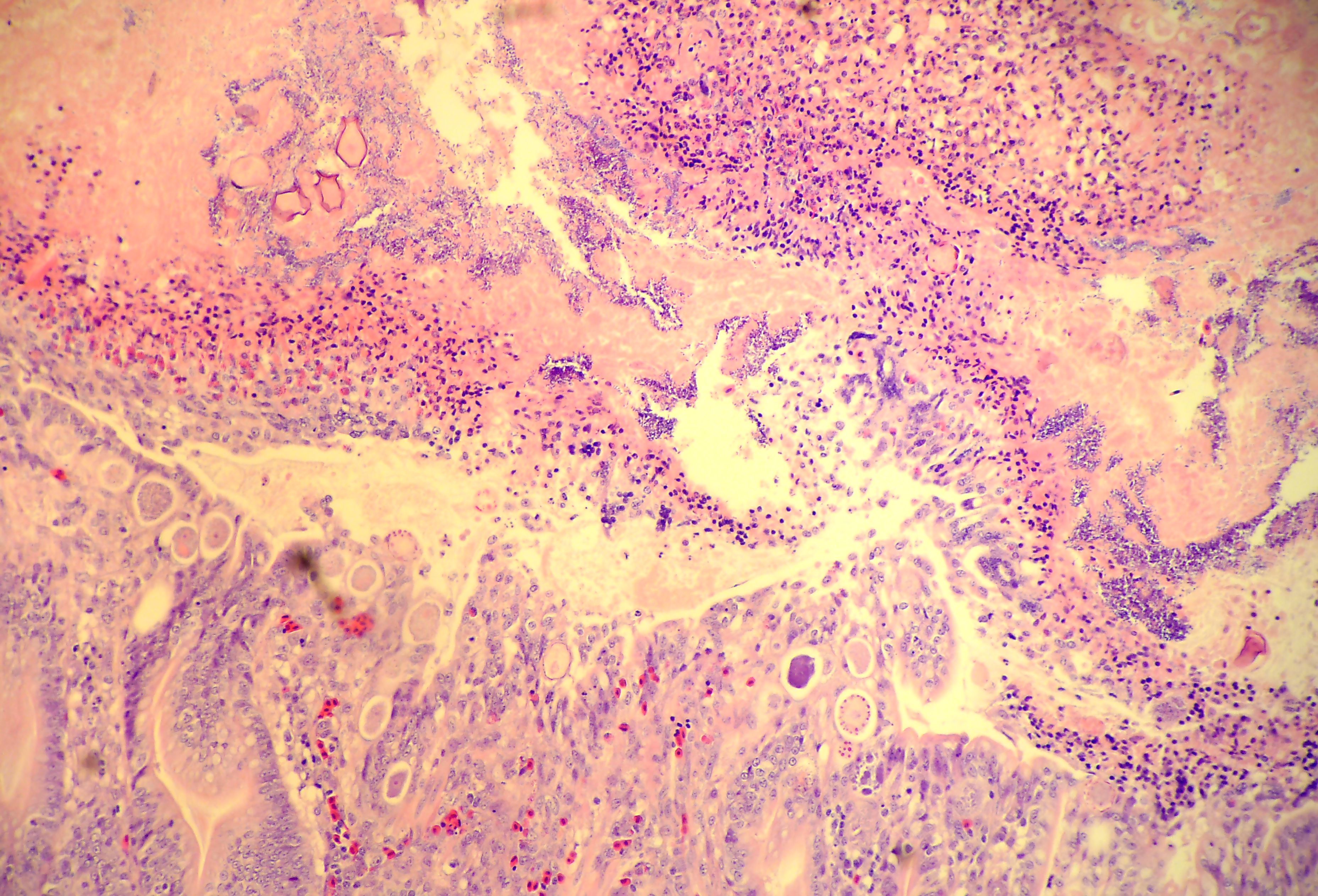
Photomicrograph depicting necrosis of the intestinal mucosa with coccidia and several rod-shaped bacteria.

Filed Under: Case Study Tagged With: Avian, broilers, center, TVMDL

Primary Sidebar

Latest Case Studies

  • Neurologic Disease due to Bovine Herpesvirus-5 (BHV-5) Infection in a 3-week-old Charolais mix calf

    February 27, 2023

  • Rare case of ocular onchocerciasis in a dog from south Texas

    January 25, 2023

  • Cerebrospinal Nematodiasis (Visceral Larval Migrans) in Birds

    September 13, 2022

  • Spironucleosis (Hexamitiasis) in Quail

    August 9, 2022

  • Endocarditis in a White-Tailed Deer Caused by Trueperella pyogenes

    August 9, 2022

Footer

For Employees

  • Employee Email
  • TVMDL Rules & Procedures
  • TVMDL Career Center
  • Emergency Alert Systems
  • AgriLife People Directory
  • AgriLife Administrative Services
  • TAMUS Single Sign On
  • Facebook

State of Texas

  • Texas.gov Portal
  • Texas Veterans Portal
  • Statewide Search
  • Texas Homeland Security
  • Risk, Fraud, & Misconduct Hotline

Policies

  • Privacy and Security Policy
  • Accessibility Policy
  • Texas A&M AgriLife
  • Texas A&M Veterinary Medical Diagnostics Laboratory
  • Texas A&M Forest Service
  • Texas A&M AgriLife Extension Service
  • Texas A&M AgriLife Research

483 Agronomy Rd
College Station, TX 77843