Achyla spp. pathogen noted in alligator hatchings
By Eric Snook, DVM, PhD
With over 800,000 tests run annually, TVMDL encounters many challenging cases. Our case study series will highlight these interesting cases to increase awareness among veterinary and diagnostic communities.
Two alligator hatchlings were presented for necropsy to the Texas A&M Veterinary Medical Diagnostic Laboratory (TVMDL) from an alligator farm. The alligator farmer noticed animals in one of his pens had started developing milky looking skin lesions over the beak and the back.
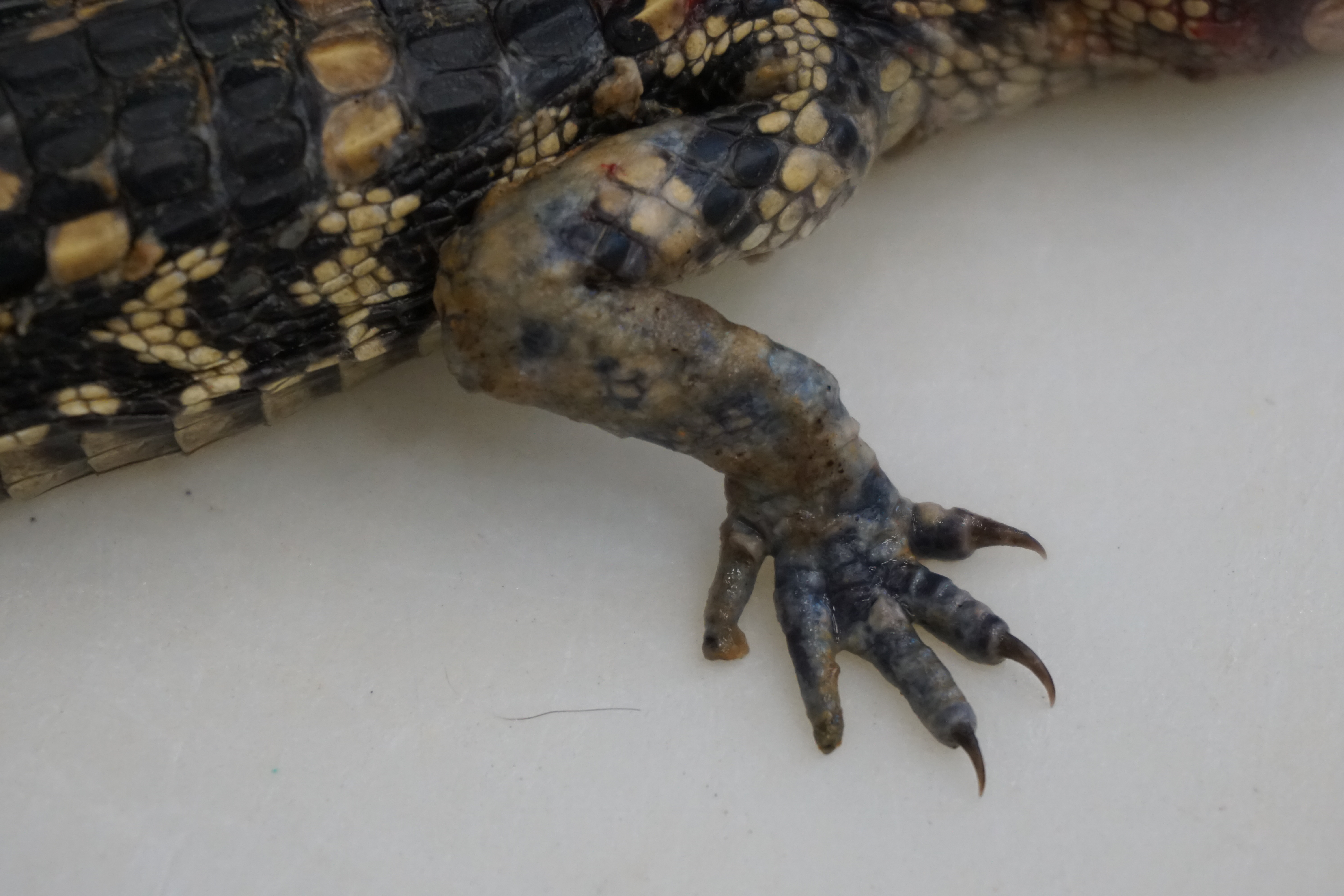
Milky lesions noted on alligator hatching
The lesions would progress, the animals would stop eating and then ultimately die. Only one pen was initially affected, but after unwittingly contaminating an adjacent pen, a second pen also developed similar lesions. Only the hatchlings were affected.
At necropsy, lesions were noted over the entire body, but were most severe over the limbs and beak. The lesions appeared to be ulcerated foci coalescing over the most severe locations. Histologically, there was a multifocal ulcerative dermatitis with numerous intralesional hyphae. Culture and DNA sequencing of the organisms identified an organism not yet reported as a pathogen in alligators: Achyla spp. Achyla spp. is an oomycete.
Oomycetes are water molds, previously classified as fungi, but now in the Kingdom Protista. Very little is understood about this organism and why these animals initially became infected, but epidemiology is currently underway to gain more understanding. Because oomycetes lack some of the key features of fungal organisms such as a chitin wall, treatment of these organisms is more challenging. The farmer in this case is implementing several treatment mechanisms to clear this organism from his farm and prevent further losses.
To learn more about this case, contact Dr. Eric Snook, veterinary pathologist at the College Station laboratory. Learn more about TVMDL and our test catalog by visiting tvmdl.tamu.edu or calling 1.888.646.5623.
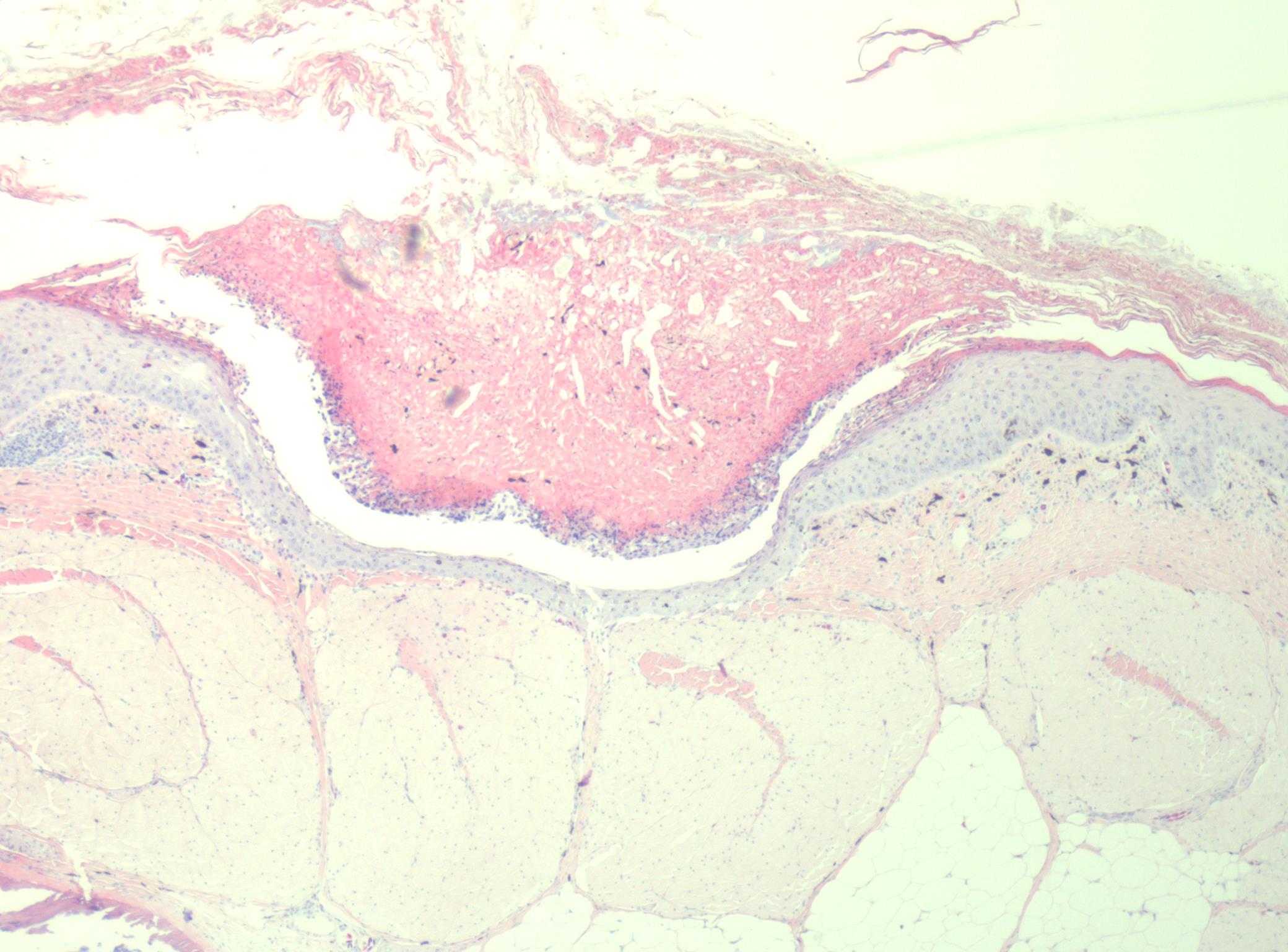
Low magnification (4x) photomicrograph of the skin over the leg demonstrating a large crust containing debris, keratin and mixed infectious agents.
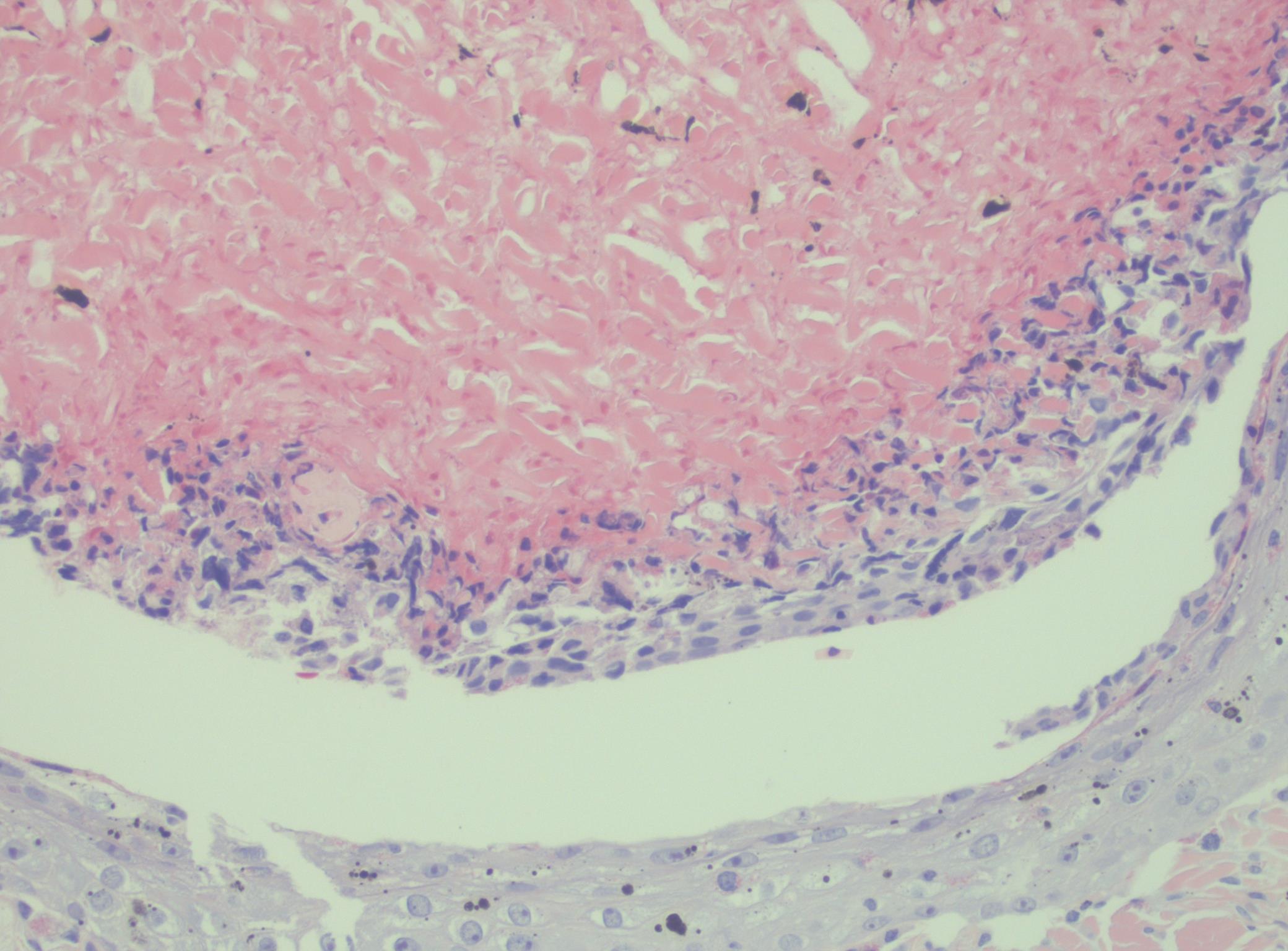
Higher magnification (20x) photomicrograph of the same area of skin demonstrating the degenerate leukocytes admixed with keratin debris and negative staining profiles of hyphae.
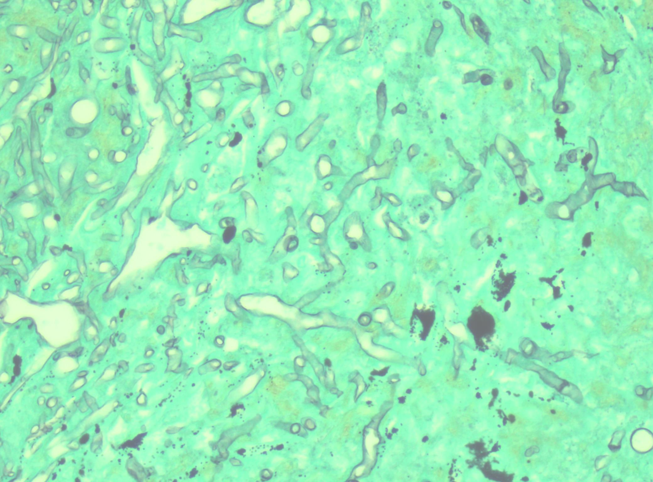
Photomicrograph at 40x with a GMS stain highlighting the abundant hyphae. The hyphae have various diameters with irregular branching and no visible septations.