• Skip to primary navigation
  • Skip to main content
  • Skip to primary sidebar
  • Skip to footer
  • Locations:
  • Canyon
  • Center
  • College Station
  • Gonzales
  • TVMDL Career Center
  • Contact Us

Texas A&M Veterinary Medical Diagnostic Laboratory

Apple App

download on the Apple App Store
download the app on Google Play

Search

Translate:

  • Find a Form
  • Deliver a Sample
    • Specimen Collection Information
    • Packaging Samples for Shipment
    • Drop Off a Sample
    • Cremation
    • Order Supplies
  • Client Portal
    • Get Test Results
    • SVA Portal
    • Pay a Bill
  • Become a Client
  • Resources
    • TVMDL Bovine Testing Guidance and Reference Material
    • Diagnostic Plans
      • Bovine Syndromic Diagnostic Plans
      • Equine Syndromic Diagnostic Plans
      • Small Ruminant Syndromic Diagnostic Plans
      • Small Animal Syndromic Diagnostic Plans
    • Education Library
    • Case Study Library
    • Electronic Reporting: QR Coding Process
    • CoreOne Resources
  • About Us
    • Locations and Hours
    • Agency Leadership
    • Speaker’s Bureau
    • Texas Pullorum-Typhoid Program
    • TVMDL Mobile app
    • Contact Us
  • Locations
    • College Station Laboratory
    • Canyon Laboratory
    • Center Laboratory
    • Gonzales Laboratory
  • Contact Us

Canine Coccidiosis

October 21, 2019 by Mallory Pfeifer

Canine Coccidiosis
Jai Guha, Mindy Borst, LVT, and Julie Piccione, DVM, MS, DACVP

An 11-year-old Corgi mix was presented to the veterinarian with intermittent diarrhea and inappetence. History noted the dog had clinical signs of gastrointestinal (GI) disease for the past few months, was current on vaccinations and was fed a raw food diet. A fecal sample was submitted to the Texas A&M Veterinary Medical Diagnostic Laboratory (TVMDL) for testing to rule out intestinal parasitism. Fecal floatation testing revealed a tremendous number of coccidia oocysts (Figure 1).

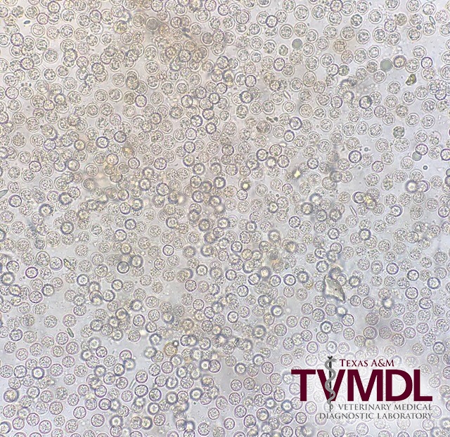
Coccidia are microscopic, single celled parasites that can infect the intestinal tract of dogs, causing diarrhea. Coccidia are typically identified based on the size and shape of the oocysts shed in feces. However, the oocysts found in this case looked similar to Hammondia heydorni, Toxoplasman gondii, and Neospora caninum. These three coccidia are morphologically similar and the oocysts are almost indistinguishable via microscopic examination due to their similar sizes (9-14 um). Real time PCR for T. gondii and N. caninum was performed on the fecal sample at TVMDL and no DNA was detected, leaving Hammondia heydorni as the only remaining identity for the parasite. Identification of the oocyst is important since T. gondii and N. caninum can cause severe disease in dogs while H. heydorniis largely considered clinically insignificant.

Hammondia heydorni is a coccidian whose definitive hosts are dogs and coyotes. Its encysted form is found in raw meats such as beef and can cause infection when consumed. Typically, the parasite does not cause harm to its definitive host; however, studies have shown that H. heydorni can cause diarrhea in immunosuppressed dogs. H. heydorni undergoes sexual reproduction in the GI tract of the definitive host, forming cysts that are excreted through the feces. The intermediate prey species (typically livestock) then ingest the cysts. Once ingested, H. heydorni attacks the prey’s muscle tissues causing atrophy and continues to reproduce inside the prey. Clinically, treatment for severe cases include sulfonamides and supportive fluid therapy. In order to prevent coccidiosis, raw meat diets should be avoided and fecal contamination should be properly disinfected.

Tremendous numbers of Hammondia heydorni oocysts seen at 400x magnification. Recovered via zinc sulfate fecal floatation.

Figure 1. Tremendous numbers of Hammondia heydorni oocysts seen at 400x magnification. Recovered via zinc sulfate fecal floatation.

To learn more about this case, contact the College Station clinical pathology section at 1.888.646.5623. For more information about TVMDL’s test offerings, visit tvmdl.tamu.edu.

References:
Schares, G., Pantchev, N., Barutzki, D., Heydorn, A. O., Bauer, C. & Conraths, F. J. (2005) Oocysts of Neospora caninum, Hammondia heydorni, Toxoplasma gondiiand Hammondia hammondiin faeces collected from dogs in Germany. International Journal for Parasitology. 35, 1525-1537.

Kahn, C. M. (2005). The Merck veterinary manual. 9th ed. / Whitehouse Station, N.J.; Great Britain: Merck & Co.

Filed Under: Case Study Tagged With: canine, clinical pathology, college station, dog, parasitology, TVMDL

Primary Sidebar

Latest Case Studies

  • Neurologic Disease due to Bovine Herpesvirus-5 (BHV-5) Infection in a 3-week-old Charolais mix calf

    February 27, 2023

  • Rare case of ocular onchocerciasis in a dog from south Texas

    January 25, 2023

  • Cerebrospinal Nematodiasis (Visceral Larval Migrans) in Birds

    September 13, 2022

  • Spironucleosis (Hexamitiasis) in Quail

    August 9, 2022

  • Endocarditis in a White-Tailed Deer Caused by Trueperella pyogenes

    August 9, 2022

Footer

For Employees

  • Employee Email
  • TVMDL Rules & Procedures
  • TVMDL Career Center
  • Emergency Alert Systems
  • AgriLife People Directory
  • AgriLife Administrative Services
  • TAMUS Single Sign On
  • Facebook

State of Texas

  • Texas.gov Portal
  • Texas Veterans Portal
  • Statewide Search
  • Texas Homeland Security
  • Risk, Fraud, & Misconduct Hotline

Policies

  • Privacy and Security Policy
  • Accessibility Policy
  • Texas A&M AgriLife
  • Texas A&M Veterinary Medical Diagnostics Laboratory
  • Texas A&M Forest Service
  • Texas A&M AgriLife Extension Service
  • Texas A&M AgriLife Research

483 Agronomy Rd
College Station, TX 77843