• Skip to primary navigation
  • Skip to main content
  • Skip to primary sidebar
  • Skip to footer
  • Locations:
  • Canyon
  • Center
  • College Station
  • Gonzales
  • TVMDL Career Center
  • Contact Us

Texas A&M Veterinary Medical Diagnostic Laboratory

Apple App

download on the Apple App Store
download the app on Google Play

Search

Translate:

  • Find a Form
  • Deliver a Sample
    • Specimen Collection Information
    • Packaging Samples for Shipment
    • Drop Off a Sample
    • Cremation
    • Order Supplies
  • Client Portal
    • Get Test Results
    • SVA Portal
    • Pay a Bill
  • Become a Client
  • Resources
    • TVMDL Bovine Testing Guidance and Reference Material
    • Diagnostic Plans
      • Bovine Syndromic Diagnostic Plans
      • Equine Syndromic Diagnostic Plans
      • Small Ruminant Syndromic Diagnostic Plans
      • Small Animal Syndromic Diagnostic Plans
    • Education Library
    • Case Study Library
    • Electronic Reporting: QR Coding Process
    • CoreOne Resources
  • About Us
    • Locations and Hours
    • Agency Leadership
    • Speaker’s Bureau
    • Texas Pullorum-Typhoid Program
    • TVMDL Mobile app
    • Contact Us
  • Locations
    • College Station Laboratory
    • Canyon Laboratory
    • Center Laboratory
    • Gonzales Laboratory
  • Contact Us

High mortality in a litter of puppies: Infection with canine herpesvirus

October 15, 2018 by Mallory Pfeifer

High mortality in a litter of puppies: Infection with canine herpesvirus
By Erin Edwards, DVM, MS, DACVP

Two Presa de Canario puppies from a litter of eight were submitted to the Texas A&M Veterinary Medical Diagnostic Laboratory (TVMDL) for necropsy. These puppies were estimated to be approximately 1 to 2 weeks old. A total of six puppies from this litter had died, amounting to a mortality rate of 75%. Some puppies were found dead with no clinical signs, while others stopped eating with death ensuing 12 to 24 hours later.

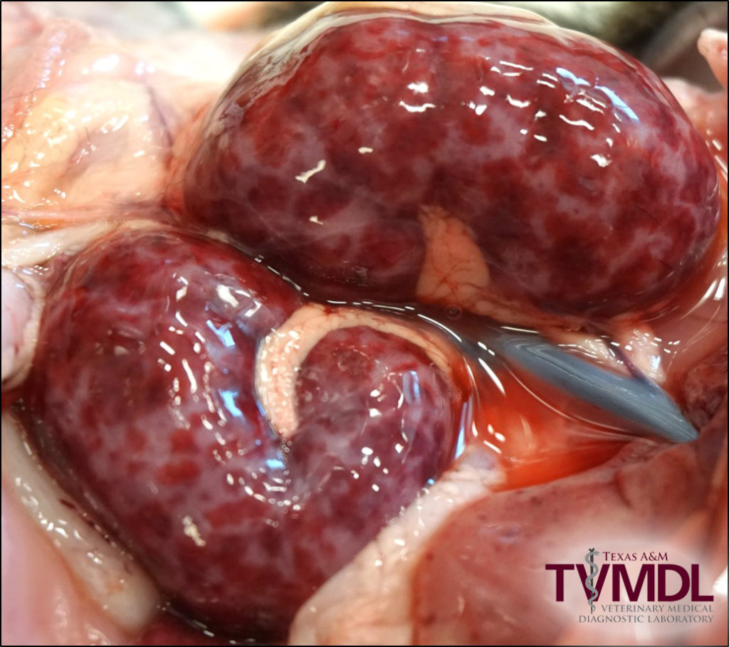
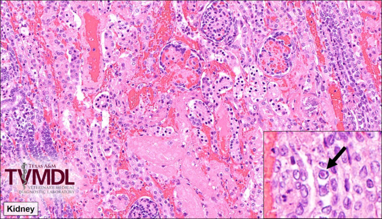
At necropsy, both puppies had similar lesions. The most striking change was multifocal to coalescing ecchymotic hemorrhages throughout the kidneys bilaterally. In one puppy, this change was so severe that nearly the entire kidney was hemorrhagic (Figure 1). Both puppies also had petechial hemorrhages throughout the liver and intestines and one puppy had a mild pneumonia. Tissues from one puppy were fixed in formalin and submitted for histopathologic examination which showed corresponding lesions of multifocal to coalescing necrohemorrhagic nephritis (Figure 2), multifocal necrohemorrhagic hepatitis, and multifocal fibrinonecrotizing pneumonia. Rare intranuclear inclusion bodies consistent with herpesvirus were seen in all affected organs (Figure 2). The macroscopic and microscopic lesions in these cases are classic for canine herpesvirus infection.

Canine herpesvirus causes systemic necrosis and hemorrhage in neonatal puppies with a high mortality rate. This disease should always be considered a major differential diagnosis in litters with multiple fatalities. Puppies can be infected in utero following infection of a naïve dam, during parturition, or following direct exposure to the virus during the first few weeks of life. Puppies aged 1-4 weeks old are the most susceptible to this disease but seldom cases are seen in puppies up to 8 weeks old. Adult dogs typically do not develop clinical symptoms from infection but can occasionally have mild upper and/or lower respiratory disease, also characterized by hemorrhage and necrosis. In puppies, the most characteristic lesion seen at necropsy is disseminated petechial to ecchymotic renal hemorrhage. Available confirmatory diagnostic tests include PCR and histopathology, though viral inclusions are sometimes difficult to find histologically.

Figure 1: Photo of kidney lesions at the time of necropsy. These kidneys have disseminated petechial to ecchymotic hemorrhages, a characteristic lesion of canine herpesvirus infection.

Figure 2: Photomicrograph depicting renal necrosis (pink area in center) and hemorrhage. Inset: Intranuclear inclusion bodies in the kidney consistent with herpesvirus.

For more information on this case, contact Dr. Erin Edwards, veterinary pathologist at the College Station laboratory.

Filed Under: Case Study Tagged With: canine, college station, histopathology, pathology, TVMDL, TVMDL-College Station

Primary Sidebar

Latest Case Studies

  • Neurologic Disease due to Bovine Herpesvirus-5 (BHV-5) Infection in a 3-week-old Charolais mix calf

    February 27, 2023

  • Rare case of ocular onchocerciasis in a dog from south Texas

    January 25, 2023

  • Cerebrospinal Nematodiasis (Visceral Larval Migrans) in Birds

    September 13, 2022

  • Spironucleosis (Hexamitiasis) in Quail

    August 9, 2022

  • Endocarditis in a White-Tailed Deer Caused by Trueperella pyogenes

    August 9, 2022

Footer

For Employees

  • Employee Email
  • TVMDL Rules & Procedures
  • TVMDL Career Center
  • Emergency Alert Systems
  • AgriLife People Directory
  • AgriLife Administrative Services
  • TAMUS Single Sign On
  • Facebook

State of Texas

  • Texas.gov Portal
  • Texas Veterans Portal
  • Statewide Search
  • Texas Homeland Security
  • Risk, Fraud, & Misconduct Hotline

Policies

  • Privacy and Security Policy
  • Accessibility Policy
  • Texas A&M AgriLife
  • Texas A&M Veterinary Medical Diagnostics Laboratory
  • Texas A&M Forest Service
  • Texas A&M AgriLife Extension Service
  • Texas A&M AgriLife Research

483 Agronomy Rd
College Station, TX 77843