• Skip to primary navigation
  • Skip to main content
  • Skip to primary sidebar
  • Skip to footer
  • Locations:
  • Canyon
  • Center
  • College Station
  • Gonzales
  • TVMDL Career Center
  • Contact Us

Texas A&M Veterinary Medical Diagnostic Laboratory

Apple App

download on the Apple App Store
download the app on Google Play

Search

Translate:

  • Find a Form
  • Deliver a Sample
    • Specimen Collection Information
    • Packaging Samples for Shipment
    • Drop Off a Sample
    • Cremation
    • Order Supplies
  • Client Portal
    • Get Test Results
    • SVA Portal
    • Pay a Bill
  • Become a Client
  • Resources
    • TVMDL Bovine Testing Guidance and Reference Material
    • Diagnostic Plans
      • Bovine Syndromic Diagnostic Plans
      • Equine Syndromic Diagnostic Plans
      • Small Ruminant Syndromic Diagnostic Plans
      • Small Animal Syndromic Diagnostic Plans
    • Education Library
    • Case Study Library
    • Electronic Reporting: QR Coding Process
    • CoreOne Resources
  • About Us
    • Locations and Hours
    • Agency Leadership
    • Speaker’s Bureau
    • Texas Pullorum-Typhoid Program
    • TVMDL Mobile app
    • Contact Us
  • Locations
    • College Station Laboratory
    • Canyon Laboratory
    • Center Laboratory
    • Gonzales Laboratory
  • Contact Us

Infectious Coryza in Chickens

November 25, 2019 by Mallory Pfeifer

Infectious Coryza in Chickens
Martin Ficken, DVM, PhD

Six 16-week-old male chickens from a flock of 500 birds were presented for necropsy at the Texas A&M Veterinary Medical Diagnostic Laboratory (TVMDL) in Gonzales. History noted one-week of swollen sinuses with minimal abnormal respiratory sounds and negligible mortality.  The incidence was relatively low.

Upon necropsy examination, each of the six birds had one of the sinuses swollen while the other sinus appeared relatively normal (Figure 1).  A thick honey-like exudate was extracted from the sinuses of three birds. Two of the six birds also had a small abscess in the left wattle.  No other lesions of note were observed.

Bacterial cultures taken from the swollen sinus of each bird were plated on sheep blood agar and MacConkey plates. A Staphylococcus aureus nurse colony was provided on the blood agar plate for supplementation. Plates were aerobically incubated at 37o C with 5% CO2 for 24 hours.  From each bird, small bacterial colonies were isolated along the S. aureus nurse colony which were identified as Avibacterium paragallinarum.

Following testing, bacterial sinusitis (infectious coryza) caused by Avibacterium paragallinarum was determined as a diagnosis.

Infectious coryza is an acute respiratory disease of chickens caused by Avibacterium paragallinarum, once known as Haemophilus paragallinarum. It causes a catarrhal inflammation of mucus membranes of the nasal passages and sinuses. Infraorbital sinuses can be distended with thick mucus. Often, there is a mild conjunctivitis and edema of the face and wattles. Uncomplicated infections rarely cause mortality; however, co-infections with other bacteria, mycoplasmas, and viruses can result in significant mortality. Once recovered, birds are carriers and can periodically shed and be a source of infection for naïve chickens.

Differential diagnoses for an acute upper respiratory infection of chickens includes fowl cholera, mycoplasmosis, ornithobacteriosis, swollen head syndrome (infectious bronchitis virus and Escherichia coli co-infection), and avitaminosis A.

For more information about this case, contact Gonzales Resident Director Dr. Martin Ficken. To learn more about TVMDL’s test offerings, visit tvmdl.tamu.edu.

Reference:
Blackall, PJ and Soriano-Vargas, E.  Infectious Coryza and Related Bacterial Infections in Diseases of Poultry, 13th edition, Wiley-Blackwell, ed. Swayne, DE et al.  pp. 859-873, 2013.


Deceased chicken with discolored beak and crown

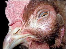
Figure 1 – Chicken with severely swollen infraorbital sinus from Avibacterium paragallinarum infection. File photograph courtesy of Dr. H. John Barnes, College of Veterinary Medicine, North Carolina State University.

Filed Under: Case Study Tagged With: Gonzales, histopathology, necropsy, pathology, TVMDL

Primary Sidebar

Latest Case Studies

  • Neurologic Disease due to Bovine Herpesvirus-5 (BHV-5) Infection in a 3-week-old Charolais mix calf

    February 27, 2023

  • Rare case of ocular onchocerciasis in a dog from south Texas

    January 25, 2023

  • Cerebrospinal Nematodiasis (Visceral Larval Migrans) in Birds

    September 13, 2022

  • Spironucleosis (Hexamitiasis) in Quail

    August 9, 2022

  • Endocarditis in a White-Tailed Deer Caused by Trueperella pyogenes

    August 9, 2022

Footer

For Employees

  • Employee Email
  • TVMDL Rules & Procedures
  • TVMDL Career Center
  • Emergency Alert Systems
  • AgriLife People Directory
  • AgriLife Administrative Services
  • TAMUS Single Sign On
  • Facebook

State of Texas

  • Texas.gov Portal
  • Texas Veterans Portal
  • Statewide Search
  • Texas Homeland Security
  • Risk, Fraud, & Misconduct Hotline

Policies

  • Privacy and Security Policy
  • Accessibility Policy
  • Texas A&M AgriLife
  • Texas A&M Veterinary Medical Diagnostics Laboratory
  • Texas A&M Forest Service
  • Texas A&M AgriLife Extension Service
  • Texas A&M AgriLife Research

483 Agronomy Rd
College Station, TX 77843