• Skip to primary navigation
  • Skip to main content
  • Skip to primary sidebar
  • Skip to footer
  • Locations:
  • Canyon
  • Center
  • College Station
  • Gonzales
  • TVMDL Career Center
  • Contact Us

Texas A&M Veterinary Medical Diagnostic Laboratory

Apple App

download on the Apple App Store
download the app on Google Play

Search

Translate:

  • Find a Form
  • Deliver a Sample
    • Specimen Collection Information
    • Packaging Samples for Shipment
    • Drop Off a Sample
    • Cremation
    • Order Supplies
  • Client Portal
    • Get Test Results
    • SVA Portal
    • Pay a Bill
  • Become a Client
  • Resources
    • TVMDL Bovine Testing Guidance and Reference Material
    • Diagnostic Plans
      • Bovine Syndromic Diagnostic Plans
      • Equine Syndromic Diagnostic Plans
      • Small Ruminant Syndromic Diagnostic Plans
      • Small Animal Syndromic Diagnostic Plans
    • Education Library
    • Case Study Library
    • Electronic Reporting: QR Coding Process
    • CoreOne Resources
  • About Us
    • Locations and Hours
    • Agency Leadership
    • Speaker’s Bureau
    • Texas Pullorum-Typhoid Program
    • TVMDL Mobile app
    • Contact Us
  • Locations
    • College Station Laboratory
    • Canyon Laboratory
    • Center Laboratory
    • Gonzales Laboratory
  • Contact Us

Visceral Mast Cell Tumor in a Cat

August 13, 2018 by Mallory Pfeifer

Visceral Mast Cell Tumor in a Cat
By Jay Hoffman, DVM, PhD

An 11 year-old, spayed female, domestic longhaired cat was submitted to the Texas A&M Veterinary Diagnostic Laboratory (TVMDL) for necropsy.  The clinical history mentioned depression with periodic vomiting for 2 to 3 days.  The animal was given medication for the vomiting and died soon afterwards.  The primary lesion observed at necropsy was marked splenomegaly; the spleen was approximately four times its normal size and was uniformly pink and firm.

Histologic evaluation of the spleen revealed that the normal splenic architecture was effaced by sheets of neoplastic round cells containing moderate amounts of finely granular amphophilic cytoplasm.  The nuclei were rounded, had finely clumped chromatin and contained one to several small nucleoli.  Mitotic figures were infrequent with up to two per 10 high powered fields.  Admixed with the neoplastic cells were scant infiltrates of eosinophils.  Similar aggregates of neoplastic cells were scattered throughout the liver and adrenal glands.  The neoplasm affecting the spleen, liver and adrenal glands was histologically compatible with a visceral mast cell tumor.  It is possible that degranulation of neoplastic mast cells caused the death of the animal.

Visceral mast cell tumors (MCTs) are a common neoplasm in cats and have been reported to cause up to 50% of all feline mast cell tumors.  Visceral MCTs most commonly affect the spleen and account for 15 to 26% of all feline splenic neoplasms.  Visceral MCTs often affect multiple organs and may involve the spleen, liver and intestines.  Clinical signs secondary to degranulation of mast cells is more common in visceral MCTs than in solitary cutaneous MCTs.  Other clinical signs include vomiting, diarrhea (with gastrointestinal involvement) and mastocytemia.  For visceral MCTs, widespread dissemination and metastasis are common.  Splenectomy of cats with splenic MCTs has been associated with median survival times of 12-19 months, regardless of metastasis.

Definitive diagnosis of visceral MCTs is straightforward by identification of sheets of neoplastic mast cells affecting visceral organs, most notably the spleen.  Special histochemical stains (Giemsa and toluidine blue) can be utilized to help identify poorly granulated neoplasms.

To learn more about this case, contact Dr. Jay Hoffman, pathology branch chief. For more information about TVMDL’s test offerings, call 1.888.646.5623 or visit tvmdl.tamu.edu.

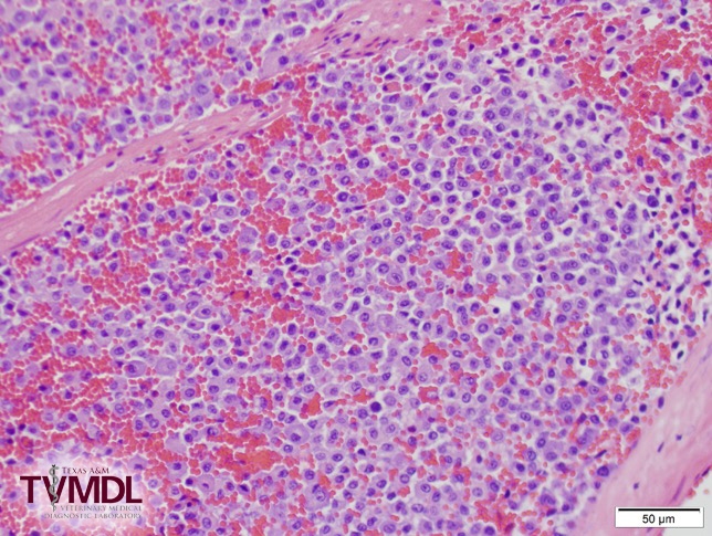
Spleen from cat. Normal architecture effaced by sheets of neoplastic mast cells.

Filed Under: Case Study Tagged With: college station, feline, histopathology, pathology, TVMDL, TVMDL-College Station

Primary Sidebar

Latest Case Studies

  • Neurologic Disease due to Bovine Herpesvirus-5 (BHV-5) Infection in a 3-week-old Charolais mix calf

    February 27, 2023

  • Rare case of ocular onchocerciasis in a dog from south Texas

    January 25, 2023

  • Cerebrospinal Nematodiasis (Visceral Larval Migrans) in Birds

    September 13, 2022

  • Spironucleosis (Hexamitiasis) in Quail

    August 9, 2022

  • Endocarditis in a White-Tailed Deer Caused by Trueperella pyogenes

    August 9, 2022

Footer

For Employees

  • Employee Email
  • TVMDL Rules & Procedures
  • TVMDL Career Center
  • Emergency Alert Systems
  • AgriLife People Directory
  • AgriLife Administrative Services
  • TAMUS Single Sign On
  • Facebook

State of Texas

  • Texas.gov Portal
  • Texas Veterans Portal
  • Statewide Search
  • Texas Homeland Security
  • Risk, Fraud, & Misconduct Hotline

Policies

  • Privacy and Security Policy
  • Accessibility Policy
  • Texas A&M AgriLife
  • Texas A&M Veterinary Medical Diagnostics Laboratory
  • Texas A&M Forest Service
  • Texas A&M AgriLife Extension Service
  • Texas A&M AgriLife Research

483 Agronomy Rd
College Station, TX 77843